MRI Scan
A Magnetic Resonance Imaging scan, or MRI scan, is a non-invasive diagnostic test that uses a magnetic field and radio waves to detect disease or injury to bones, as well as soft tissues inside your body, like your heart and blood vessels, or to identify areas of the brain affected by stroke.
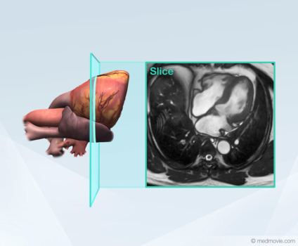
A cardiac MRI scan is a kind of MRI scan that shows how well blood flows to your heart muscle tissue and through the chambers of your heart. If needed, a contrast dye may be used to help show certain structures, like blood vessels, more clearly. The contrast dye is injected into a vein in your arm and travels to your heart.
Additionally, a medication that mimics the effects of exercise on blood vessels is often used to help detect disease or injury. The details and effects of any contrast dye or other medication will be discussed by your healthcare team.
MRI scans do not use x-ray radiation. Instead, MRI scans use a powerful magnetic field to create a series of cross-sectional images, or “slices”, which can be stacked together to create a three-dimensional model of the structures inside of your body. Although this magnetic field cannot be felt, it can be heard. Ear protection is often used for patient comfort.
The information from an MRI scan helps your physician choose an appropriate treatment. Ask your healthcare team for more details.
Visit One Heart Cardiology for more information.