Pulmonary Valve Stenosis
The pulmonary valve is located between the right ventricle and the large blood vessel to the lungs which is called the pulmonary artery. Pulmonary Valve Stenosis is a condition in which the pulmonary valve leaflets are thickened and fused, making it harder for blood to flow from the right ventricle to the lungs. Pressure rises in the right ventricle in order to pump blood through the narrowed valve. When the pressure build-up is significant, the muscle of the right ventricle becomes thickened, known as hypertrophy, and this can eventually lead to muscle damage.
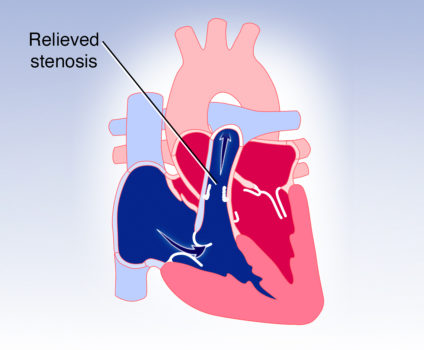
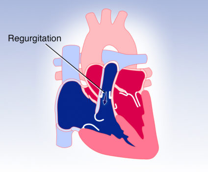
A significantly narrowed valve can be opened by balloon dilation. This procedure is called a transcatheter balloon valvuloplasty. A balloon catheter is threaded from the large vein in the groin into the right ventricle and through the narrowed pulmonary valve. The balloon on the end of the catheter is then inflated to stretch, or tear, the pulmonary valve leaflets. Once the catheter is removed, blood can flow more easily through the valve and the stenosis is relieved. This procedure is very effective for relieving the narrowing. Because the balloon valvuloplasty often tears the pulmonary valve leaflets, they may not close tightly.This causes leakage of blood backward into the ventricle, also known as insufficiency or regurgitation. Later in life patients may require a valve replacement if the insufficiency or regurgitation becomes severe.
Visit Medmovie.com Website for more information.