

In the early stages of fetal development, two aortic arches come from the heart, ascend upward and then descend behind the heart merging together to become a single aorta. As the heart develops normally, the right-sided arch disappears, leaving the left-sided arch to ascend upward and continue to the descending aorta behind the heart. The normally developed left-sided aorta lies in front of the trachea, or breathing tube, and esophagus, or swallowing tube.
A vascular ring is a defect where the arch vessels encircle the breathing and swallowing tubes.This is caused by abnormal development of the aortic arches. If both arches stay open it is called a double aortic arch. The two arches surround the breathing and swallowing tubes and may cause narrowing or compression of these structures and lead to breathing and/or feeding difficulties. In another form of a vascular ring, the left-sided arch disappears, and the right-sided arch stays open. If this is associated with an abnormal origin of the blood vessel that goes to the left arm and a left-sided ductus arteriosus, a vascular ring is formed.
Surgery can be performed in symptomatic patients to divide part of the aorta or other vascular structures in order to eliminate the compression of the breathing and swallowing tubes. For a double aortic arch, the left arch is typically divided along with the ductal ligament. For a vascular ring formed by a right aortic arch with a left-sided ductal ligament, division of the ductal ligament is performed.
©2024 Medmovie.com. All rights reserved. Medmovie.com creates and licenses medical illustrations and animations for educational use. Our goal is to increase your understanding of medical terminology and help communication between patients, caregiver and healthcare professionals. The content in the Media Library is for your information and education purposes only. The Media Library is not a substitute for professional medical advice, diagnosis or treatment for specific medical conditions.
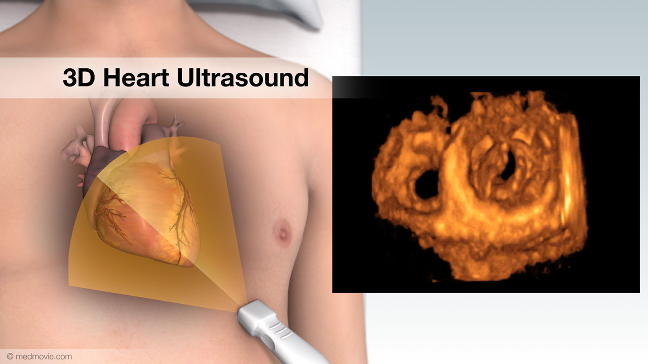 3D Heart UltrasoundA 3D heart ultrasound, also known as a 3D echocardiogram, is a diagnostic test that creates 3D videos of the heart.…
 Arterial Switch OperationTransposition of the great arteries (TGA) is a congenital heart defect in which the aorta, which normally arises from…
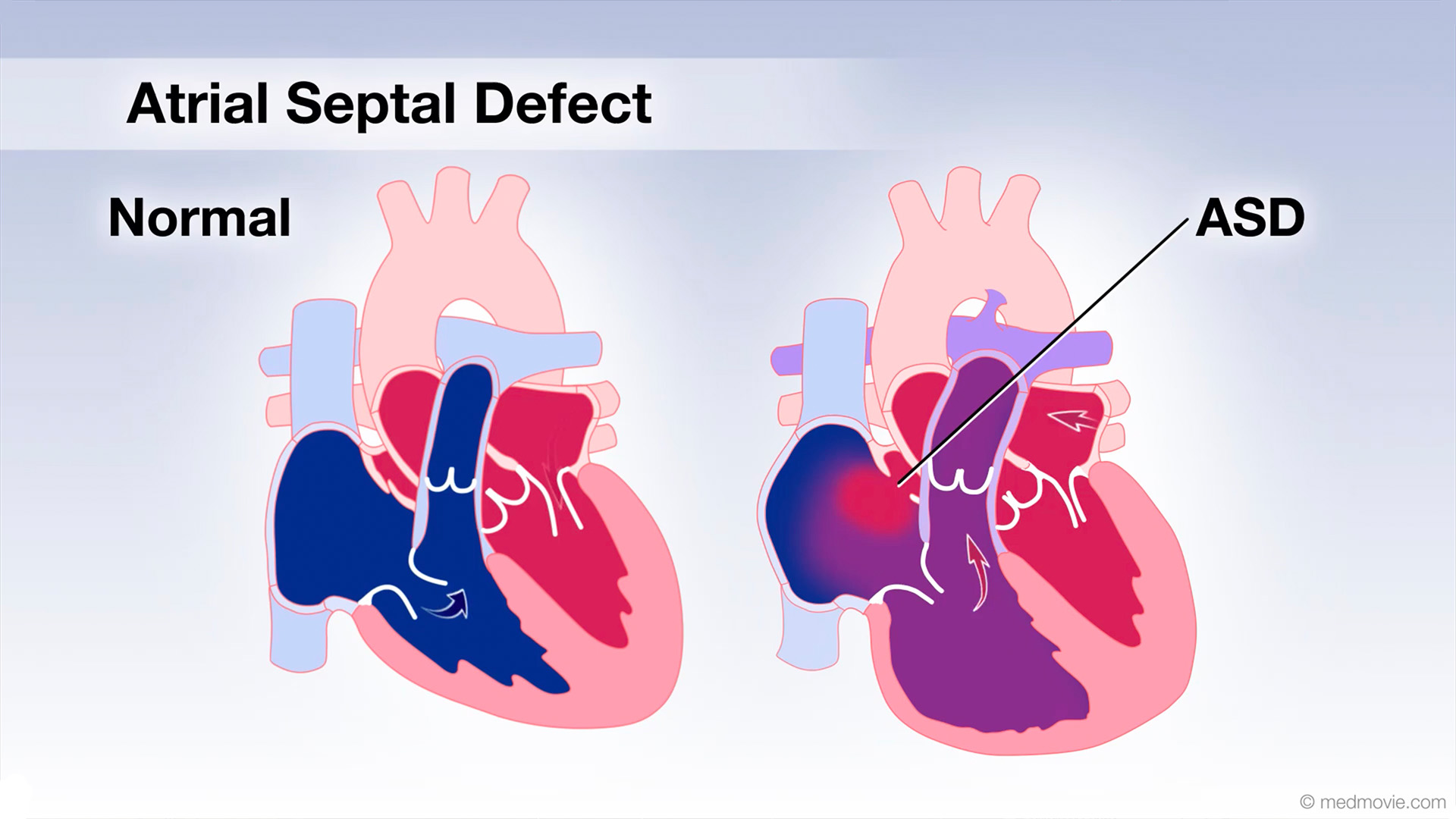 Atrial Septal DefectAn atrial septal defect, or ASD, is a hole in the wall that separates the upper two chambers of the heart, known as the…
 ASD ClosureSecundum atrial septal defect, or ASD, is a hole in the atrial septum that separates the atria. This allows oxygen-poor…
 AV CanalThe atrioventricular canal, or AV canal, is the structure that forms the center of the heart. During normal fetal…
 Balloon SeptostomyA Balloon Atrial Septostomy (or Rashkind procedure) is a procedure that is used to create an opening in the wall…
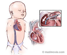 Balloon Septostomy BabyA Balloon Atrial Septostomy (or Rashkind procedure) is a procedure that is used to create an opening in the wall…
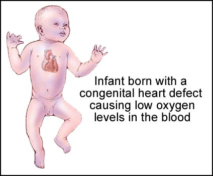 Blue BabyBlue baby is condition in which the skin of an infant appears to be blue due to low oxygen levels in the blood. This is…
 Blalock-Taussig ClassicalA Blalock-Taussig shunt is a surgical procedure that helps improve oxygenation of the blood. It is used in cases of…
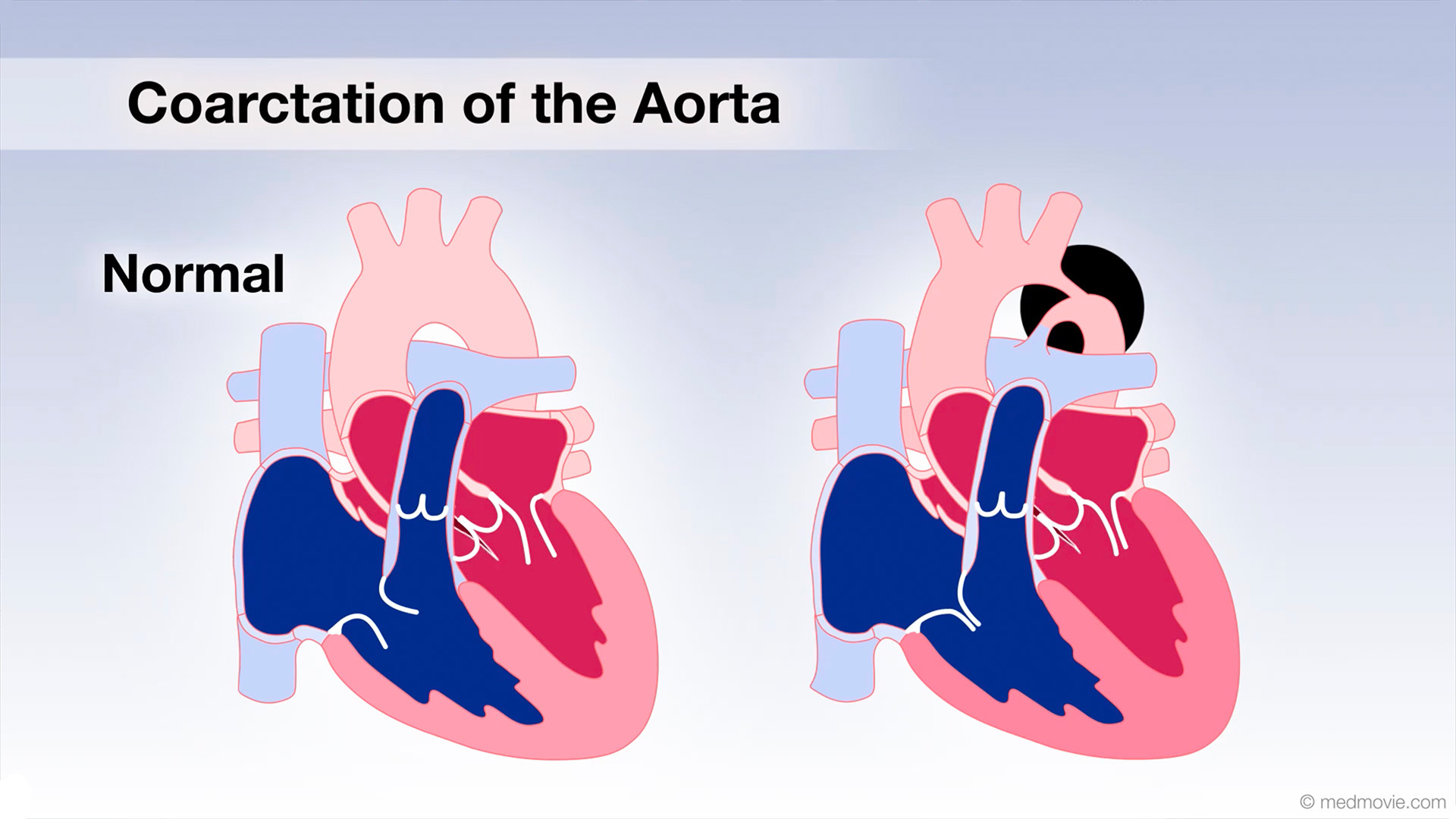 Coarctation of AortaCoarctation of the Aorta is a congenital defect in which the upper part of the descending aorta is severely narrowed or…
 DiureticsDiuretics (often called “water pills”) are drugs that cause the body to rid itself of excess fluids and sodium through…
 Doppler UltrasoundDoppler ultrasound is a test that uses high-frequency sound waves to detect the speed and direction of blood flow in…
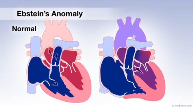 Ebstein's AnomalyThe tricuspid valve is the valve between the right atrium and the right ventricle, and in Ebstein’s anomaly it is…
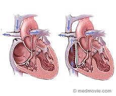 Fontan ProcedureA Fontan procedure is a series of surgical steps that is used to correct tricuspid atresia. Tricuspid atresia is a…
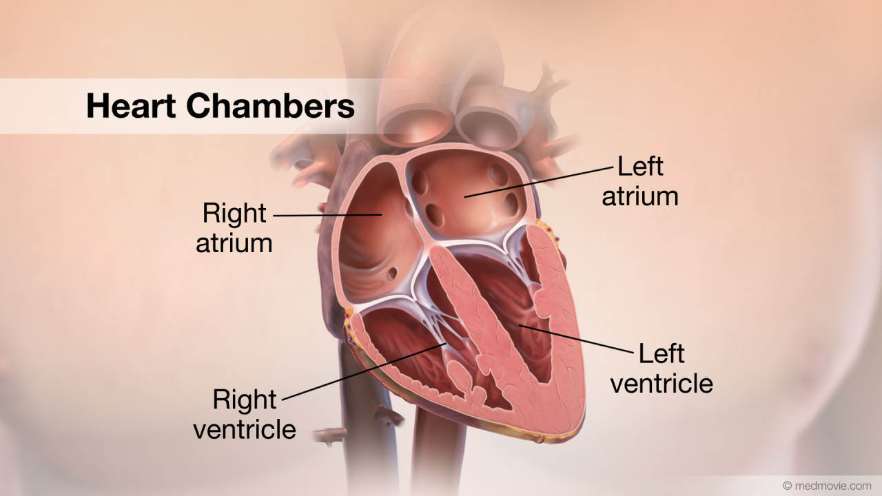 Heart ChambersThe heart is divided into four chambers. The two upper chambers are called the atria. The right atrium and left atrium…
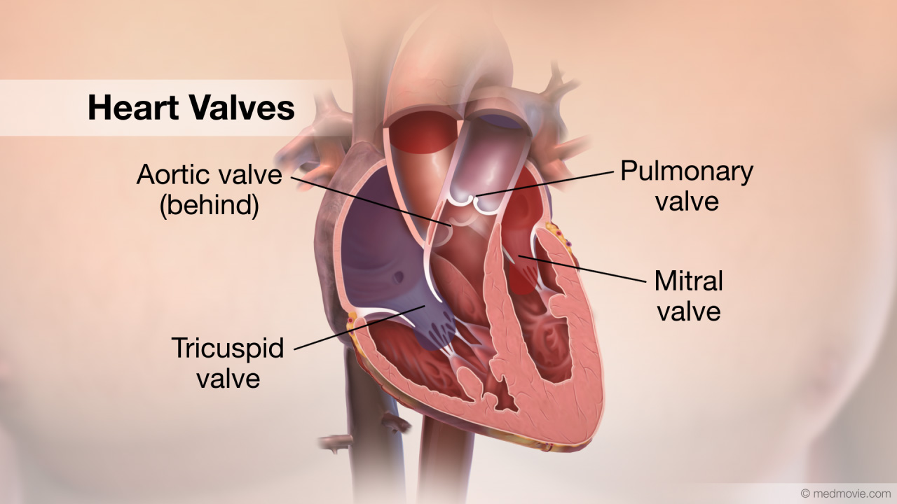 Heart ValvesThe heart has four valves. The mitral valve, the tricuspid valve, the pulmonary valve and the aortic valve. The mitral…
 Hypertrophic CardiomyopathyThe normal heart has strong muscular walls that pump blood out of the heart.
Hypertrophic cardiomyopathy is a…
 Hypoplastic Left Heart SyndromeHypoplastic left heart syndrome is the result of underdevelopment of the left heart structures and includes a tiny, or…
 Blalock-Taussig ModifA Blalock-Taussig shunt is a surgical procedure that helps improve oxygenation of the blood. It is used in cases of…
 Norwood ProcedureA Norwood procedure is used to correct a type of congenital heart defect called hypoplastic left heart syndrome. This…
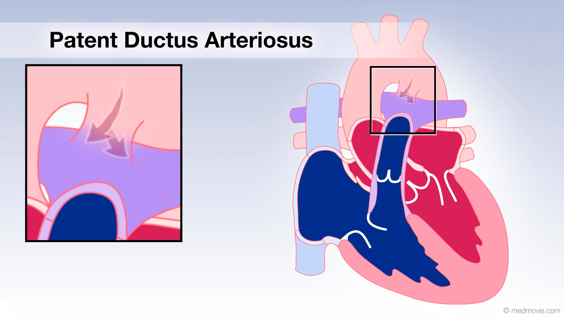 Patent Ductus Arter.The ductus arteriosus is a blood vessel that connects the blood vessel going to the lungs, known as the pulmonary…
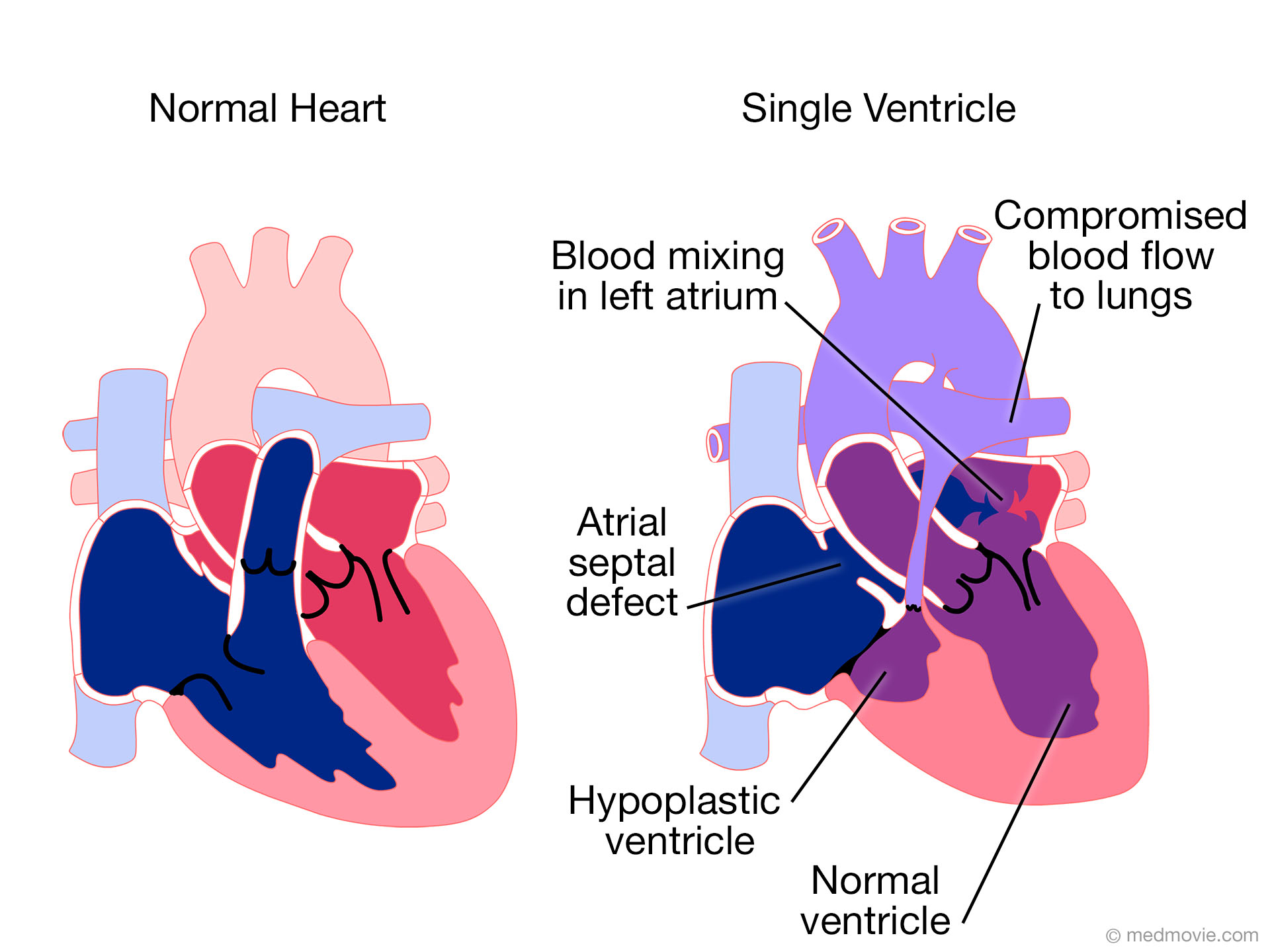 Single VentricleSingle ventricle is a congenital defect in which one ventricle of the heart is abnormally small (hypoplastic). The…
 Tetrology of FallotTetrology of Fallot is characterized by several malformations of the heart. These include: ventricular septal defect…
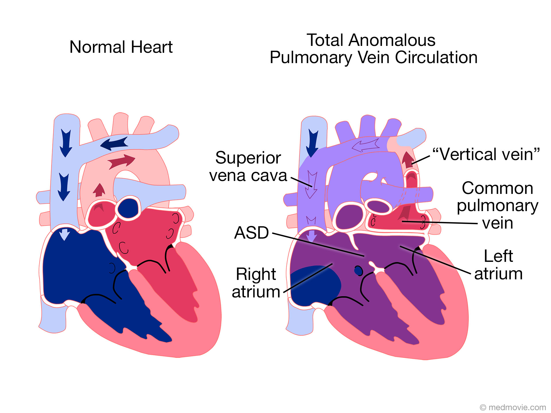 TAPVCTotal anomalous venous connections, or TAPVC, is a congenital malformation in which the pulmonary veins join to form a…
 Transp. of Great Arter.D-Transposition (shown in this animation) of the great arteries (TGA) is a congenital heart defect in which the aorta,…
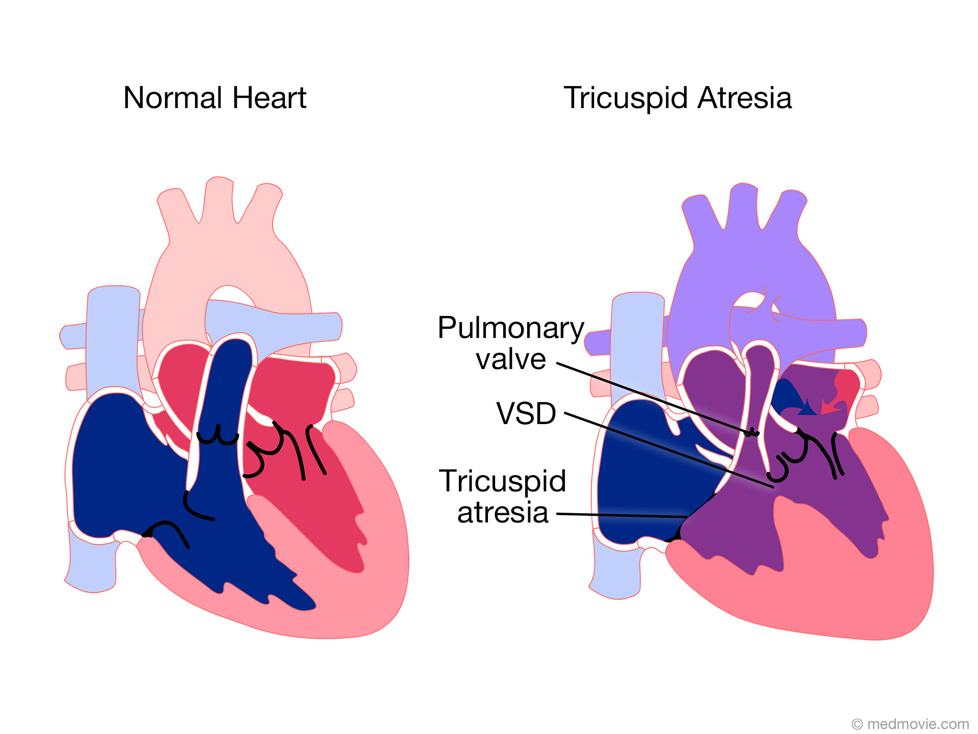 Tricuspid AtresiaTricuspid atresia with is a congenital defect in which the tricuspid valve that lies between the right atrium of the…
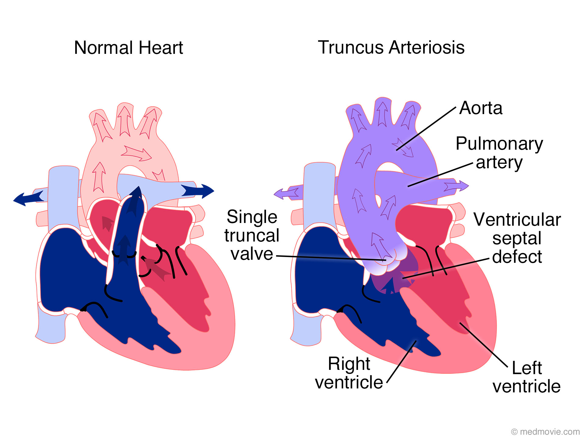 Truncus ArteriosisTruncus arteriosus is a congenital defect in which the two major arteries (the aorta and the pulmonary artery) fail to…
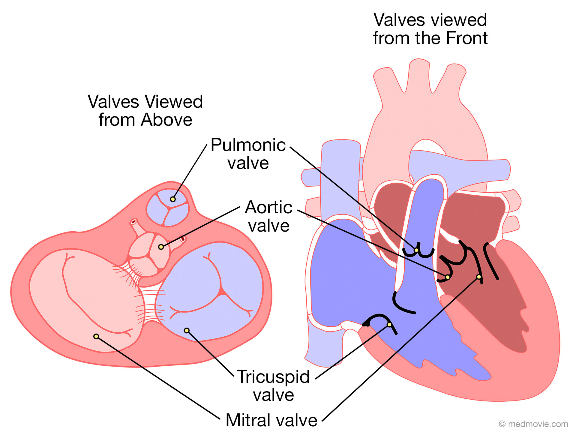 Valves- 2 ViewsThe heart has four valves that regulate blood flow through the heart. The mitral valve and the tricuspid valve…
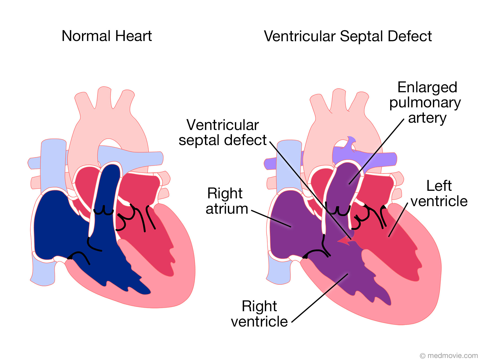 Ventric. Septal DefectVentricular septal defect, or VSD, is a hole in the septum that separates the right and left ventricles of the heart.…
 WPW SyndromeWolff-Parkinson White Syndrome (WPW) is a condition in which the heart beats too fast due to abnormal, extra electrical…
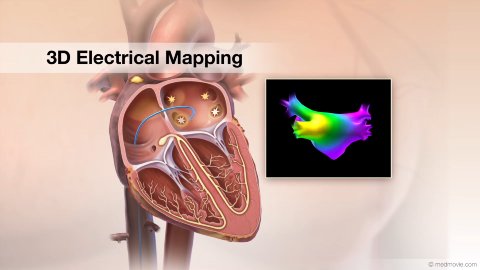 3D Electrical MappingElectrical mapping of the heart is a procedure that is used to diagnose the origins of arrhythmias. This procedure uses…
 3D Heart UltrasoundA 3D heart ultrasound, also known as a 3D echocardiogram, is a diagnostic test that creates 3D videos of the heart.…
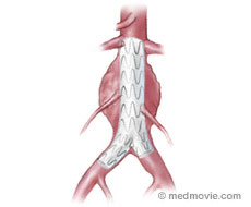 Abdominal Aortic Aneurysm Stent GraftAn aortic endovascular stent graft is used to treat abdominal aortic aneurysms and is an alternative to a surgical…
 AAA SurgeryAortic aneurysm that require repair can be repaired surgically. Surgery usually involves placing a synthetic tube into…
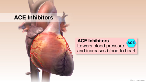 ACE InhibitorsACE Inhibitors (Angiotensin-Converting Enzyme) Inhibitors are drugs used to treat high blood pressure and heart failure…
 Active Fixation LeadAn active fixation lead is a pacemaker or implantable defibrillator lead that is fixed to the inside surface of the…
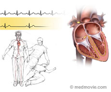 Adams-Stokes DiseaseAdams-Stokes disease is a temporary condition caused by a heart arrhythmia. Adams-Stokes is caused inadequate blood…
 Aldosterone AntagonistsAldosterone Antagonists are drugs that act as diuretics (water pills). Diuretics cause the kidneys to remove water from…
 Alpha BlockersAlpha Blockers are drugs that treat a variety of conditions, such as high blood pressure, benign prostatic hyperplasia…
 AnginaAngina is the term used to describe a broad range of symptoms due to blockages in the coronary arteries that reduce…
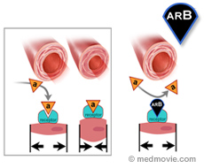 ARBsAngiotensin II Receptor Blockers (ARBs) are drugs that are used to treat high blood pressure and heart failure. They…
 ABI TestThe ankle-brachial index (ABI) test is a painless exam that compares the blood pressure in your feet to the blood…
 AntiarrhythmicsAntiarrhythmics are drugs that help control the heart arrhythmias. An arrhythmia is a condition in which abnormal…
 AnticoagulantsAnticoagulants are drugs that decrease the ability of the blood to clot (coagulate). Blood clotting is caused by a…
 AntihypertensivesAntihypertensives are drugs commonly prescribed to help lower blood pressure when appropriate diet and regular physical…
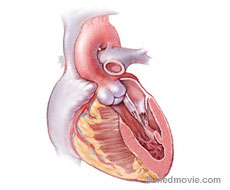 AortaThe aorta is the largest artery in the body. Oxygenated blood is pumped from the left ventricle of the heart into the…
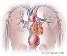 Aortic AneurysmAn aortic aneurysm is an enlargement or buldging of the aorta. The aorta is the large artery into which blood is pumped…
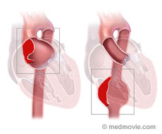 Aortic AneurysmAn aortic aneurysm is an enlargement or bulging of the aorta. The aorta is the large artery into which blood is pumped…
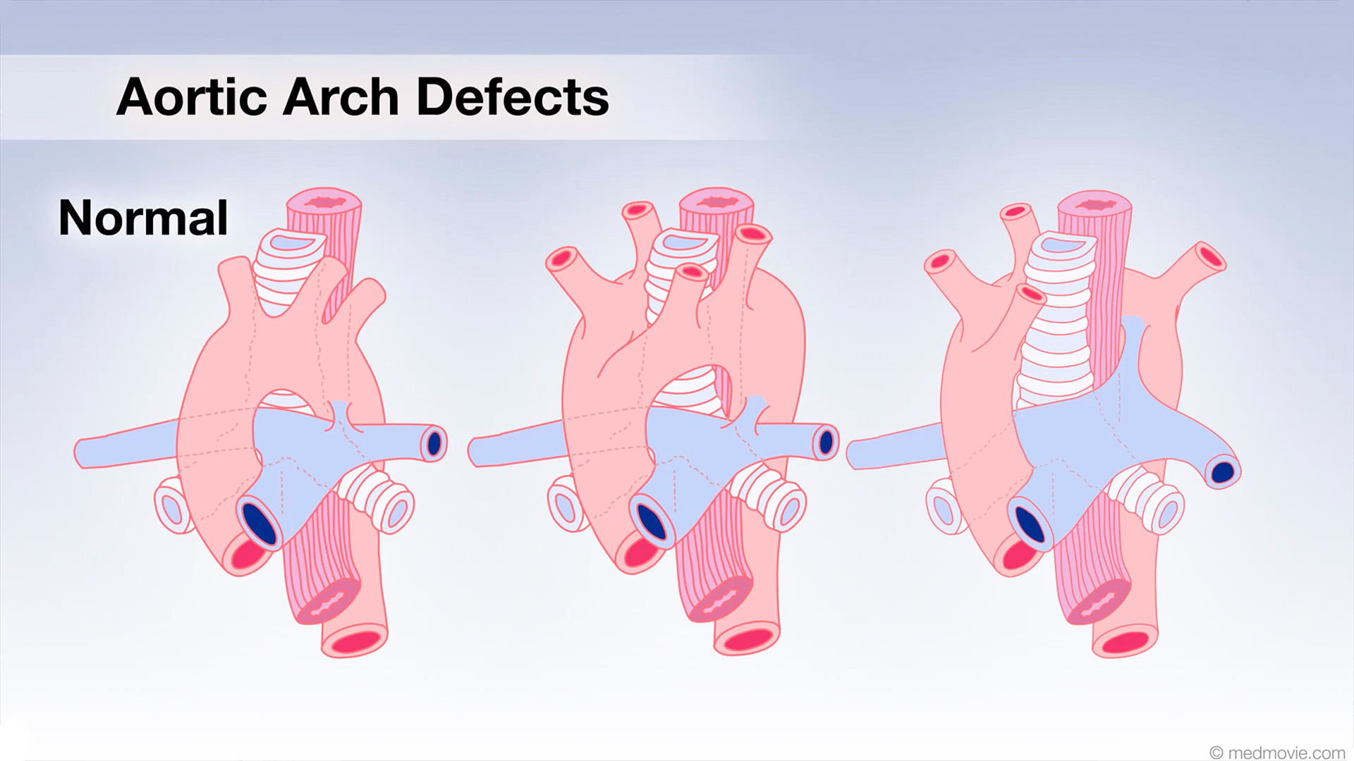 Aortic Arch DefectsIn the early stages of fetal development, two aortic arches come from the heart, ascend upward and then descend behind…
 Aortic ValveThe aortic valve is one of four valves that control blood flow through the heart, keeping blood flowing in a forward…
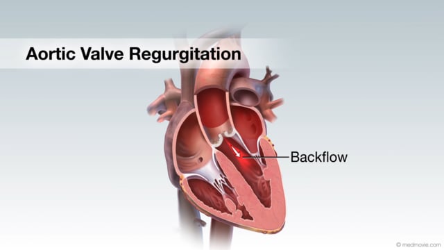 Aortic Valve RegurgitationAortic valve regurgitation results when the aortic valve, which sits between the left ventricle and the aorta, cannot…
 Aortic Valve StenosisThe aortic valve is located between your heart’s main pumping chamber (left ventricle) and the major artery (aorta)…
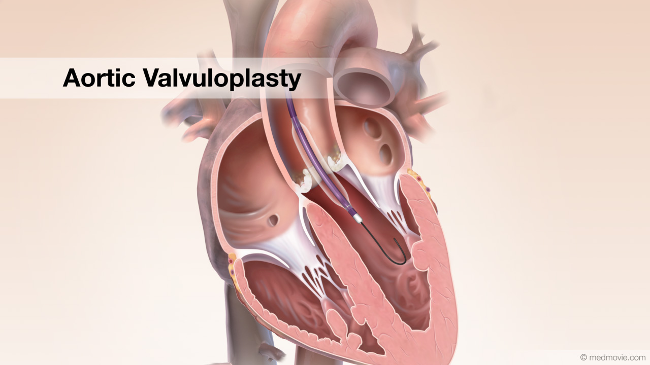 Aortic ValvuloplastyStenotic aortic valve disease can require a procedure called aortic valvuloplasty. Calcific aortic stenosis is shown…
 ApexThe apex is the pointed tip of the heart. It is located on the lower portion of the heart (left ventricle).
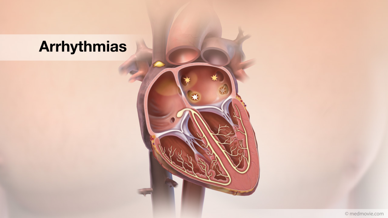 ArrhythmiasThe heartbeat is controlled by the electrical system of the heart. This system is made up of several sequences that…
 Arterial AnatomyArteries are part of the circulatory system of the body. Arteries carry oxygenated blood from the heart to the body and…
 Arterial GraftsSections of arteries in the body can be used as grafts to reroute blood around arterial blockage that cut off blood…
 Arterial Switch OperationTransposition of the great arteries (TGA) is a congenital heart defect in which the aorta, which normally arises from…
 AsprinAspirin has been shown to help prevent heart attack and stroke. Aspirin works by affecting the blood clotting process.…
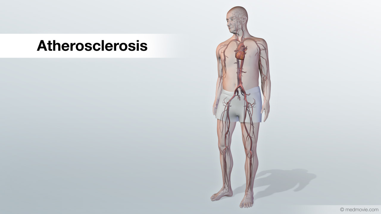 AtherosclerosisAtherosclerosis is a disease that affects the arteries, which carry blood to the organs of the body.
Normal arteries…
 Atherosclerosis ComplicationsAtherosclerosis is a disease where fatty material called plaque builds up in the inner lining of arteries. Plaque…
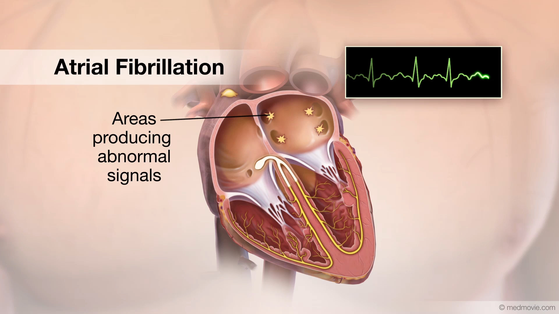 Atrial FibrillationThe heartbeat is controlled by the electrical system of the heart. This system is made up of several parts that tell…
 Atrial Fibrillation AblationAtrial fibrillation is a heart arrhythmia in which abnormal electrical signals begin in the atria (top chambers) Atrial…
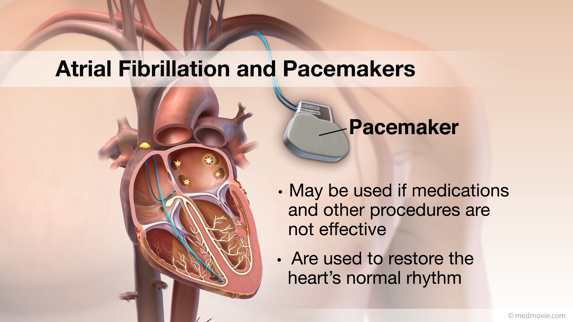 Atrial Fibrillation PacemakersAtrial fibrillation is a heart arrhythmia in which abnormal electrical signals begin in the atria (top chambers) of the…
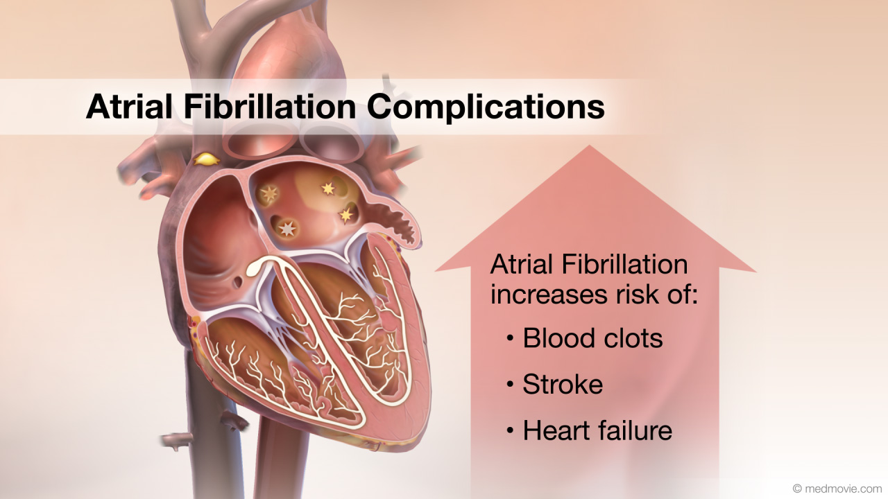 Atrial Fibrillation ComplicationsAtrial fibrillation (AF) is a heart arrhythmia in which abnormal electrical signals begin in the atria (top chambers)…
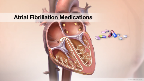 Atrial Fibrillation MedicationsAtrial Fibrillation is a heart arrhythmia in which abnormal electrical signals begin in the atria (or top chambers) of…
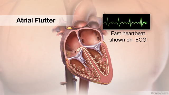 Atrial FlutterThe heartbeat is controlled by the electrical system of the heart. This system is made of several parts that tell the…
 Atrial Flutter AblationAtrial flutter ablation is a procedure used to correct atrial flutter by destroying small areas of tissue to block…
 Atrial Septal DefectAn atrial septal defect, or ASD, is a hole in the wall that separates the upper two chambers of the heart, known as the…
 ASD ClosureSecundum atrial septal defect, or ASD, is a hole in the atrial septum that separates the atria. This allows oxygen-poor…
 Atrial TachycardiaThe heartbeat is controlled by the electrical system of the heart. This system is made up of several parts that tell…
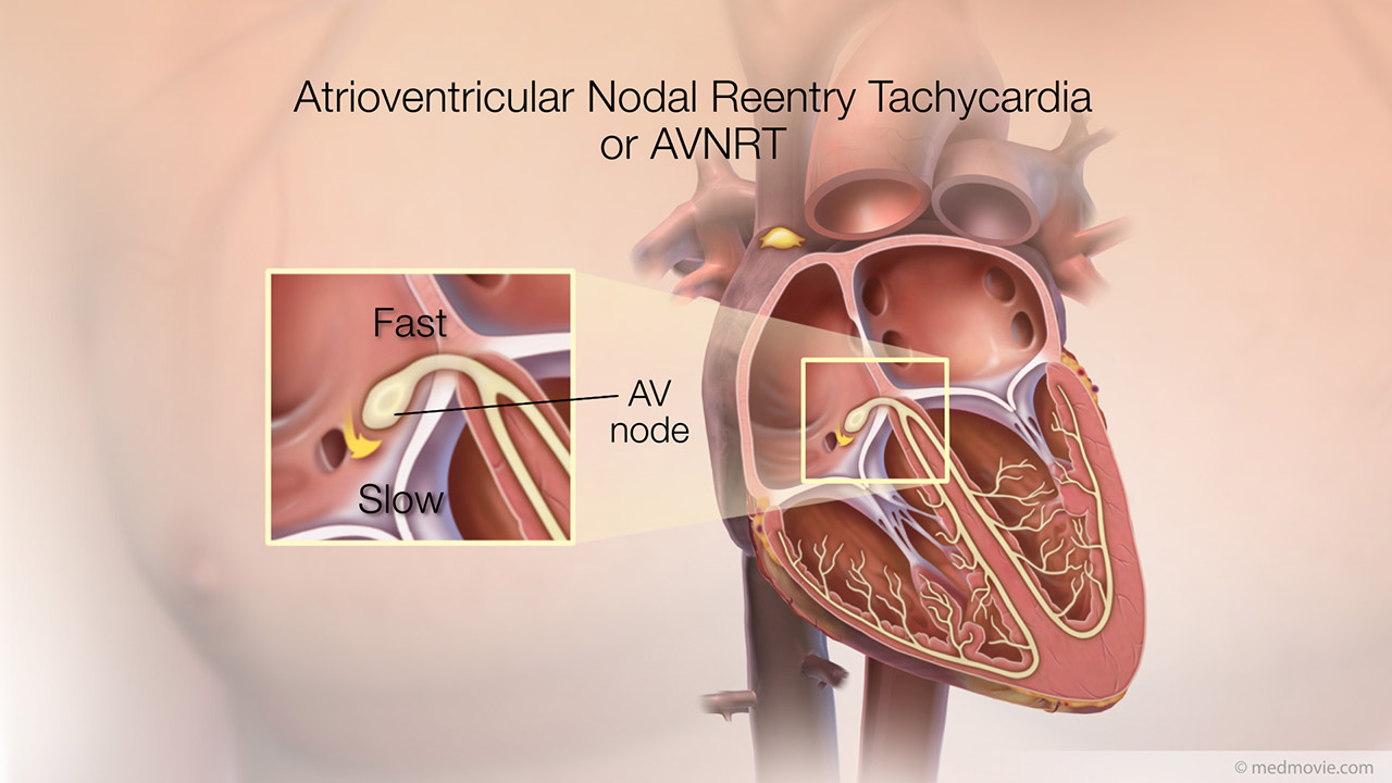 AVNRTThe heartbeat is controlled by the electrical system of the heart. This system is made up of several parts that tell…
 AVRTThe heartbeat is controlled by the electrical system of the heart. This system is made up of several parts that tell…
 AV CanalThe atrioventricular canal, or AV canal, is the structure that forms the center of the heart. During normal fetal…
 AV Node AblationAV node ablation is a procedure used to correct irregular heartbeats by destroying the tissue within the AV node. AV…
 Bacterial EndocarditisBacterial endocarditis is an infection of the inner lining (endocardium) of the heart’s chambers or valves. It is…
 Balloon SeptostomyA Balloon Atrial Septostomy (or Rashkind procedure) is a procedure that is used to create an opening in the wall…
 Balloon Septostomy BabyA Balloon Atrial Septostomy (or Rashkind procedure) is a procedure that is used to create an opening in the wall…
 Balloon CatheterA balloon dilation catheter is an instrument used to treat atherosclerotic narrowing of arteries. It consists of a…
 Beta BlockersBeta Blockers are drugs that slow the heart rate, decrease cardiac output, lessen the force with which the heart muscle…
 Blood ClotA blood clot is a normal reaction of the body that occurs if a vessel wall is injured. Blood clots are formed by a…
 Blood PressureBlood Pressure is the pressure of the blood exerted against the walls of the arteries. Optimal blood pressure is less…
 Blue BabyBlue baby is condition in which the skin of an infant appears to be blue due to low oxygen levels in the blood. This is…
 BradycardiaThe heartbeat is controlled by the electrical system of the heart. This system is made of several parts that tell the…
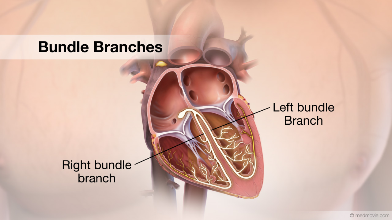 Bundle BranchesThe bundle branches are a part of the electrical system of the heart.
The electrical system controls the heartbeat…
 Bundle of HisThe bundle of His is a part of the electrical system of the heart. It is a collection of cells that carry electrical…
 Calcium Channel BlockersCalcium Channel Blockers are drugs that block the movement of calcium into heart and blood vessel muscle cells, which…
 CapillariesCapillaries are part of the circulatory system. They are tiny vessels that connect the smallest arteries and veins.…
 Cardiac CatheterizationCardiac catheterization refers to a procedure used to diagnose and treat certain heart conditions.
To initiate a…
 CRT DeviceA cardiac resynchronization therapy (CRT) device is a battery-powered device that sends electrical signals to your…
 Cardiac TamponadeCardiac tamponade is a condition in which the heart is “strangled” by a buildup of fluid within the sac (the…
 Carotid AngiographyA carotid angiogram is a test that uses X-rays to show narrowing or blockage in the carotid arteries. The carotid…
 Carotid StentingA carotid stent is a small wire mesh tube that is used to hold open a carotid artery that has been narrowed by artery…
 Carotid ArteriesThe carotid arteries are the main source of blood supply to the head and neck. They also provide the main blood supply…
 Carotid Art. DiseaseCarotid artery disease is a condition that causes the narrowing of the carotid artery by the formation of…
 Carotid Artery SurgeryCarotid artery surgery, also known as carotid endarterectomy, is a procedure that removes fatty deposits (plaque) that…
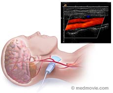 Carotid IMTCarotid IMT provides your doctor with moving images of your carotid artery and takes excellent pictures that will help…
 Catheter AblationThe electrical system of the heart controls each heartbeat. Electrical impulses generated by special tissue (nodes)…
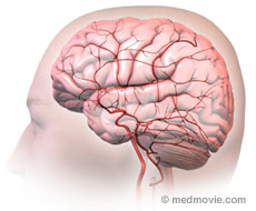 Cerebrovascular Sys.The cerebrovascular system is a term for the blood vessels that carry blood to and from the brain. The arteries (shown…
 Cholesterol-HDL & LDCholesterol is a fatty substance present throughout the body. Cholesterol comes form two sources; it is made in the…
 Cholesterol SourcesThe liver produces about 1,000 mg of cholesterol a day. Another 200 to 500 mg can come from food.
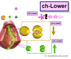 Cholesterol-Lowering DrugsCholesterol-Lowering Drugs help reduce the “bad” cholesterol (LDL), increase the “good” cholesterol (HDL) and reduce…
 Kidney DiseaseChronic kidney disease, also known as renal disease, is progressive loss of kidney function.
Stages of kidney…
 Blalock-Taussig ClassicalA Blalock-Taussig shunt is a surgical procedure that helps improve oxygenation of the blood. It is used in cases of…
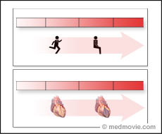 Classification of Cardiovascular DiseaseClassification of Functional Capacity and Objective Assessment is an in-patient screening criteria for cardiac disease…
 Coarctation of AortaCoarctation of the Aorta is a congenital defect in which the upper part of the descending aorta is severely narrowed or…
 CommissurotomyThe valves of the heart can be affected by several diseases that result in failure to open properly, thereby…
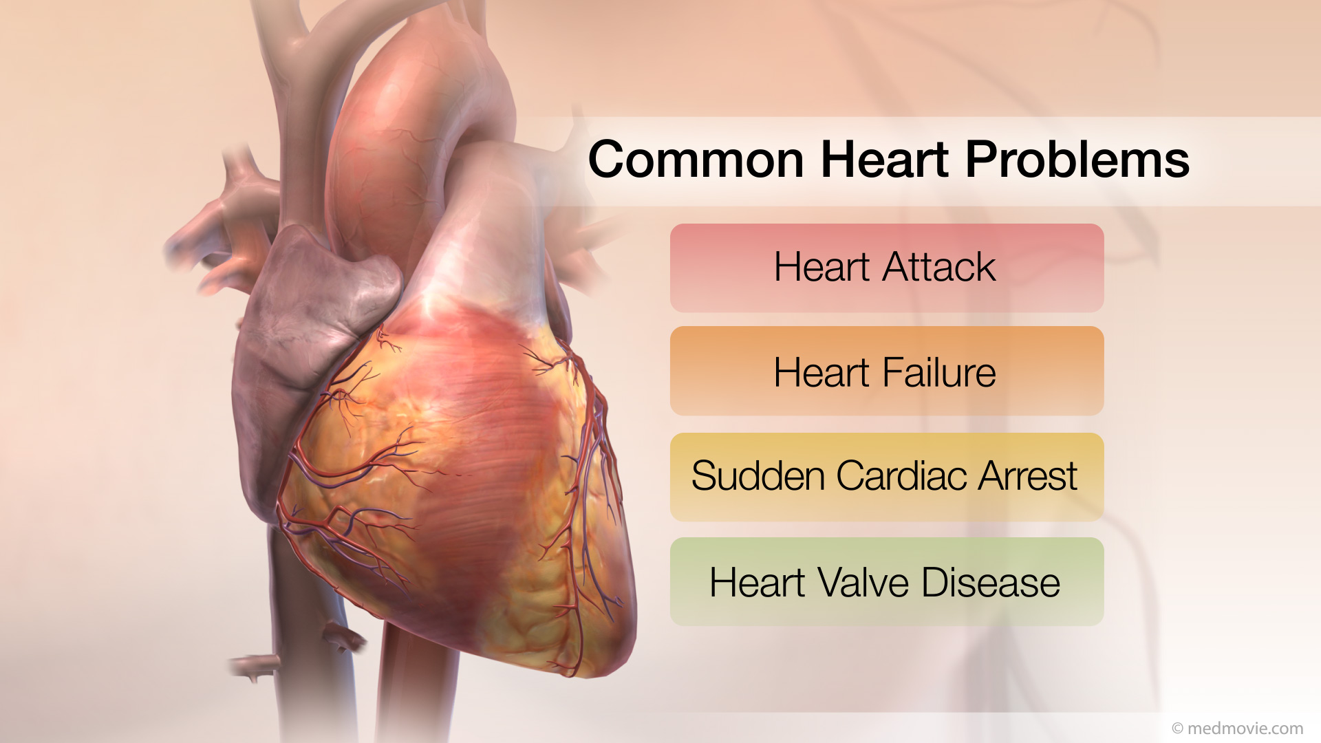 Common Heart ProblemsThe arteries, muscle, conductive tissue and valves of the heart work together to keep it beating normally. These four…
 Communicating with Your Healthcare ProviderThe American Heart Association encourages you to actively discuss all aspects of your health. Talking with your…
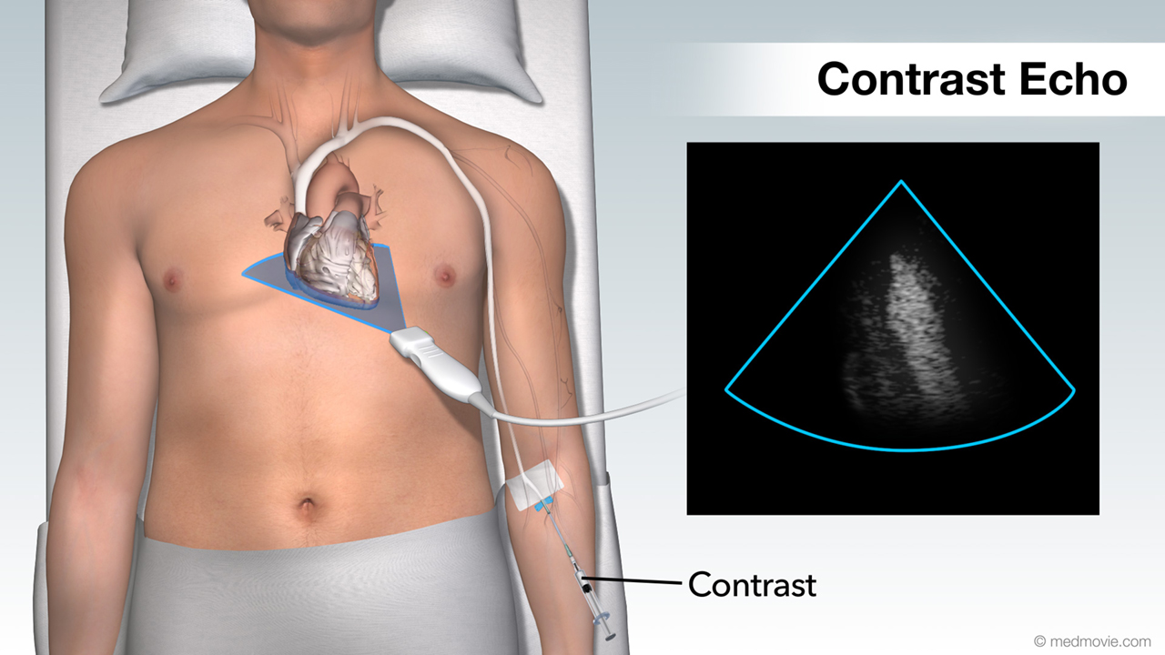 Contrast EchoHeart Ultrasound provides your doctor with moving images of your heart and takes excellent pictures that will help your…
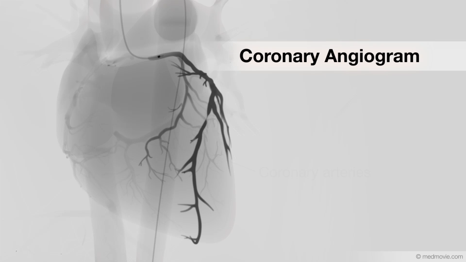 Coronary AngiogramA coronary angiogram is a test that uses x-ray imaging and contrast dye to view the coronary arteries, the blood…
 Coronary AngioplastyCoronary angioplasty (PTCA) is a procedure that opens up narrowed or blocked segments of the arteries that supply blood…
 Coronary ArteriesThe coronary arteries are the blood vessels that supply blood to the heart muscle. They branch off of the aorta at its…
 Coronary Bypass GraftCoronary artery bypass graft surgery (CABG) creates new pathways around narrowed or blocked coronary arteries so that…
 Coronary Art. DiseaseCoronary artery disease is a condition in which the coronary arteries are narrowed by deposits called plaques.
The…
 Coronary Artery SpasmA coronary artery spasm is a sudden, temporary contraction of muscle fibers within the walls of the coronary artery.…
 Coronary AtherectomyCoronary atherectomy is a procedure utilized to restore normal blood flow through a blocked vessel by removing the…
 Coronary CTO TherapyCoronary chronic total occlusion therapy refers to one of several procedures that can be used to treat coronary…
 Coronary StentA Coronary Stent is a small expandable tube shaped device used to open narrowed coronary arteries that have interrupted…
 CPRCPR, or cardiopulmonary resuscitation, is a technique of mainting air exchange in the lungs and blood flow in the…
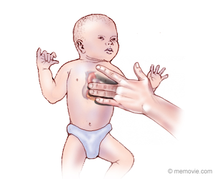 CPR: InfantInfant CPR or cardiopulmonary resuscitation is a technique that can be used revive an infant whose heart has stopped…
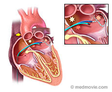 CryotherapyCryotherapy is a procedure that can be used to treat arrhythmias in the heart. This procedure uses a special catheter…
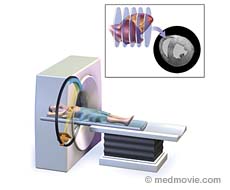 CT ScanA CT scan is a diagnostic test that uses X-rays to allow images of the body to be made. CT stands for “Computed…
 Diabetes ComplicationsDiabetes is a disease that affects greater than 20 million people in the United States. The main feature of diabetes is…
 Diabetes Types 1 and 2Diabetes is a condition caused by the body’s inability to process sugar and relates to a lack of insulin production or…
 Dilated CardiomyopathyDilated cardiomyopathy is a term used to describe a condition of the heart in which one or more of the chambers of the…
 DiureticsDiuretics (often called “water pills”) are drugs that cause the body to rid itself of excess fluids and sodium through…
 Doppler UltrasoundDoppler ultrasound is a test that uses high-frequency sound waves to detect the speed and direction of blood flow in…
 Ebstein's AnomalyThe tricuspid valve is the valve between the right atrium and the right ventricle, and in Ebstein’s anomaly it is…
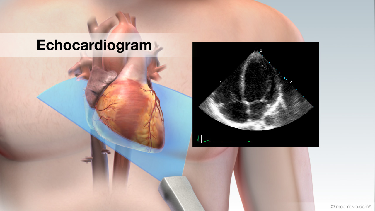 EchocardiogramAn echocardiogram, or cardiac ultrasound, is a diagnostic test that uses sound waves to view structures of the beating…
 Ejection FractionDuring each heartbeat, the heart contracts and relaxes. The ventricles are the pumping chambers of the heart. In…
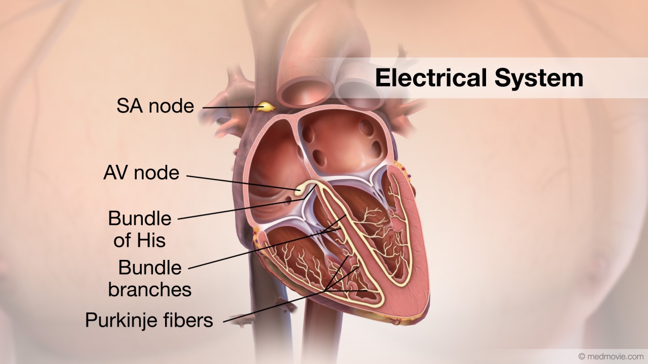 Electrical SystemThe purpose of the electrical system of the heart is to coordinate the pumping of the four chambers of the heart and to…
 Heartbeat - ElectricHeart muscle cells generate electrical signals that are conducted through the heart by special fibers which stimulate…
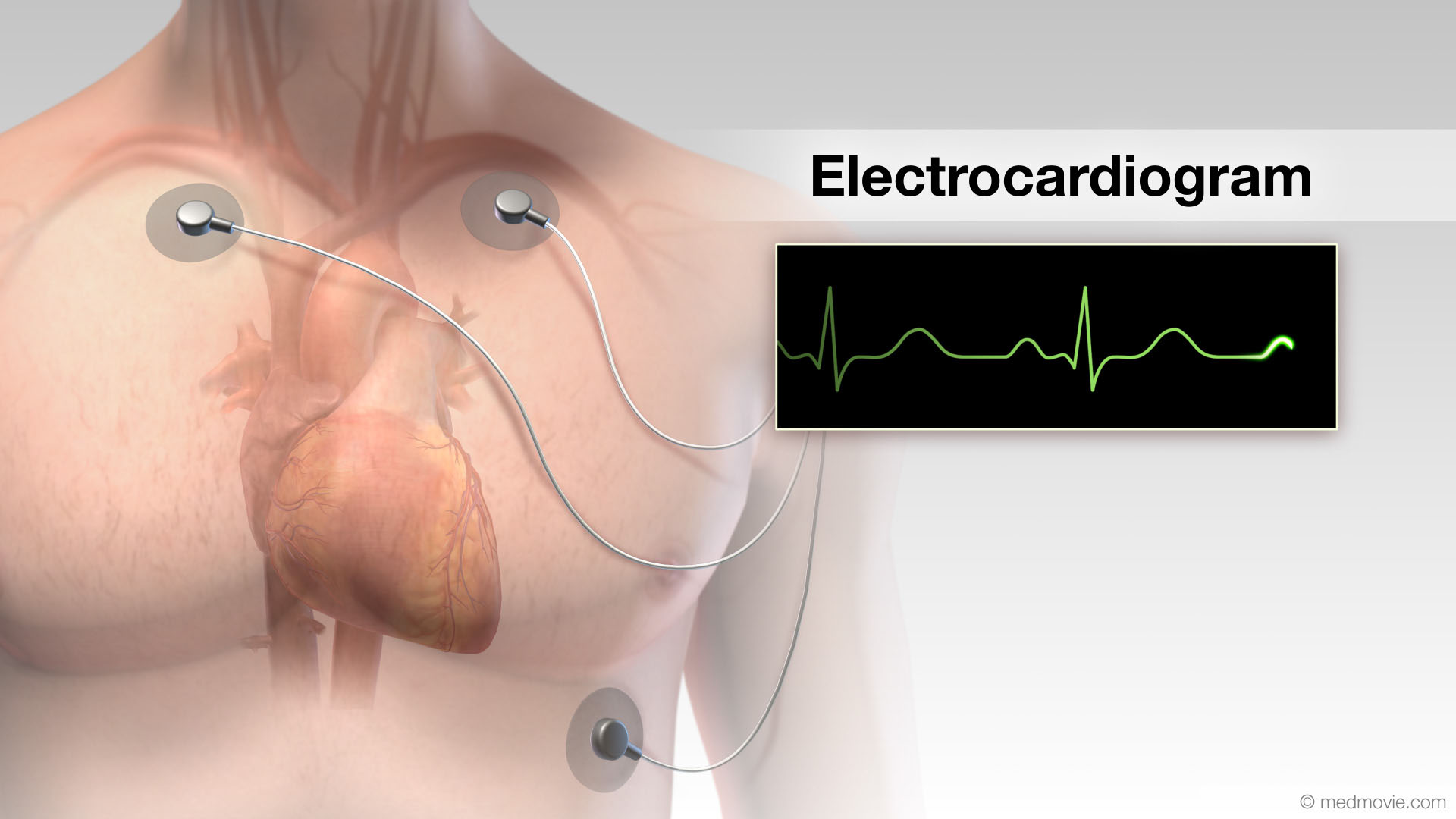 ElectrocardiogramThe electrocardiogram or ECG, records the electrical activity of the heart. It is used to determine if the heart has an…
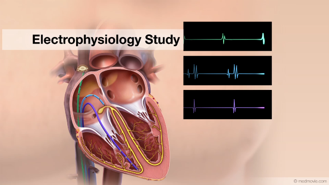 EP StudyAn electrophysiology study is performed to evaluate the electrical activity of the heart. The purpose of the electrical…
 Endovascular GraftsAn aortic endovascular stent graft is used to treat abdominal aortic aneurysms and is an alternative to a surgical…
 Exercise Stress TestAn exercise stress test is a diagnostic test in which a person walks on a treadmill or pedals a stationary bicycle…
 External DefibrillatorAn external defibrillator is a machine that is used to deliver an electrical shock to the heart to restore a normal…
 Femoral Artery StentA femoral stent is a small wire mesh tube that is used to hold open a femoral artery that has been narrowed by artery…
 Focal Atrial TachycardiaThe heartbeat is controlled by the electrical system of the heart. This system is made up of several parts that tell…
 Focal Ventricular TachycardiaThe heartbeat is controlled by the electrical system of the heart. This system is made up of several parts that tell…
 Fontan ProcedureA Fontan procedure is a series of surgical steps that is used to correct tricuspid atresia. Tricuspid atresia is a…
 Healthy EatingHealthy eating decreases the risk of heart disease and is good for the entire cardiovascular system. There are three…
 Heart and LungsThe heart can be thought of as the main pump within the circulation system. The right side of the heart collects blood…
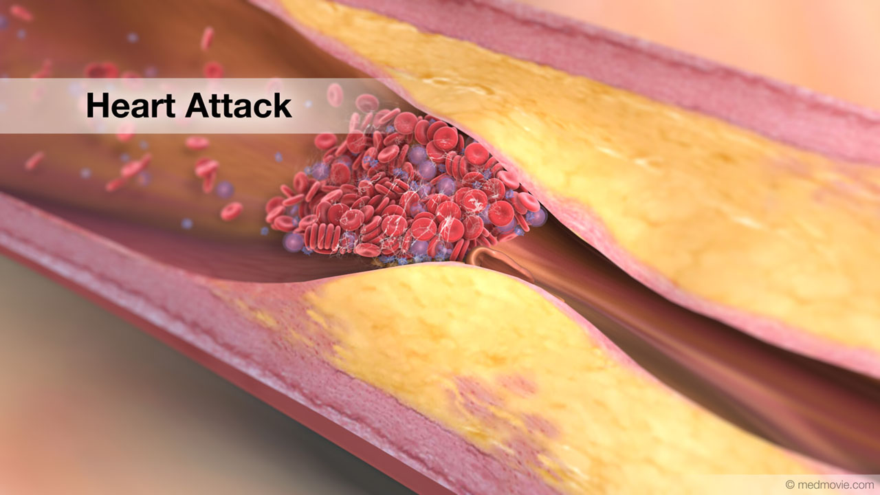 Heart AttackA heart attack happens when blood flow to an area of heart muscle suddenly becomes blocked and an area of heart muscle…
 Heart BlockHeart block is an arrhythmia in which electrical signals traveling from the atria to the ventricles are stopped or…
 Heart ChambersThe heart is divided into four chambers. The two upper chambers are called the atria. The right atrium and left atrium…
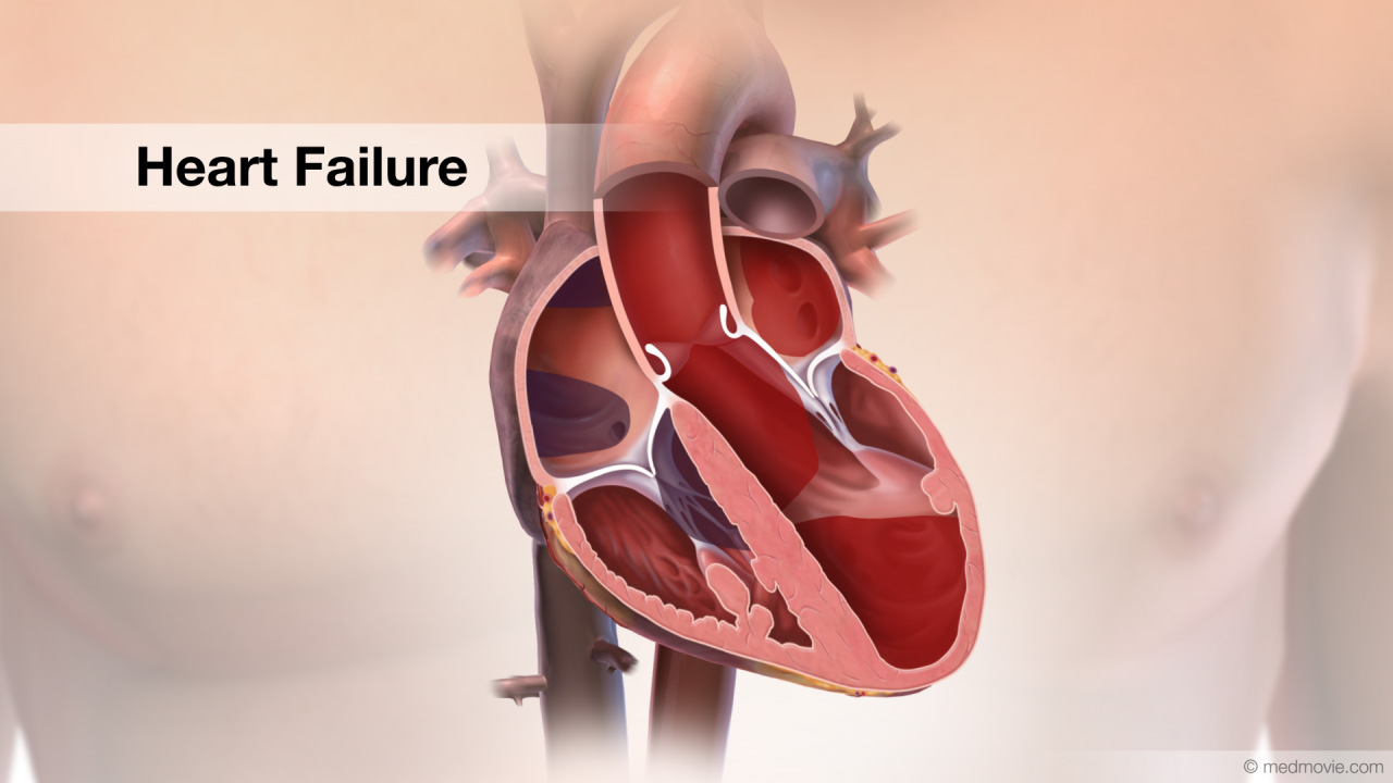 Heart FailureThe normal heart has strong muscular walls that pump blood out of the heart. Heart failure most often results from a…
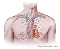 Heart LocationThe heart is located within the rib cage, under and slightly to the left of the breastbone (sternum). It approximately…
 Heart Lung BypassA heart-lung -or cardiopulmonary bypass - machine is used during heart surgery to deliver oxygenated blood to the body…
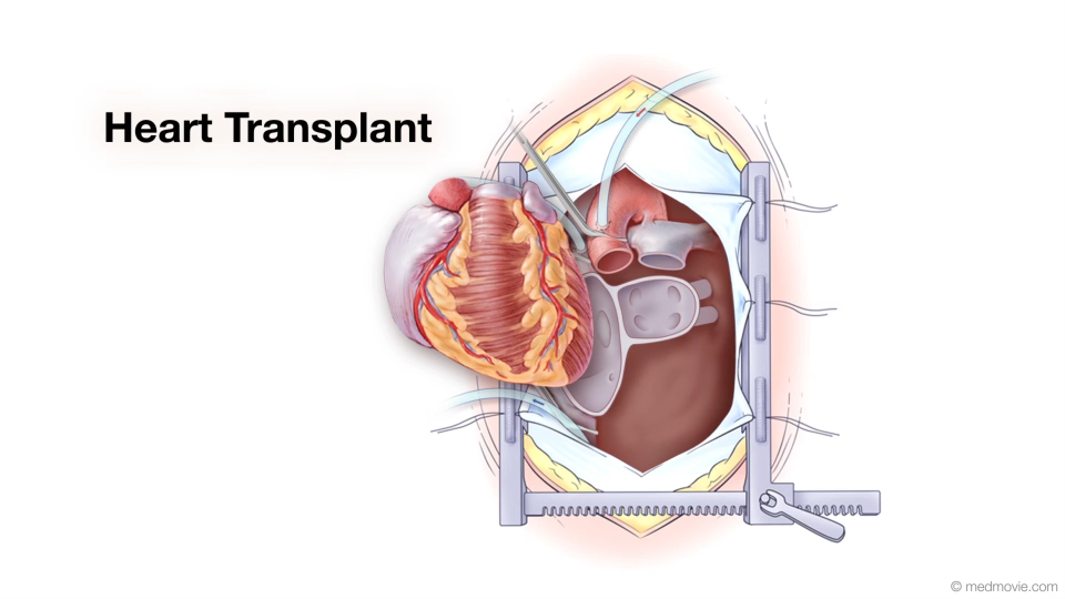 Heart TransplantA heart transplant is a surgical procedure that replaces a failing heart with a healthy donor heart. Physicians make a…
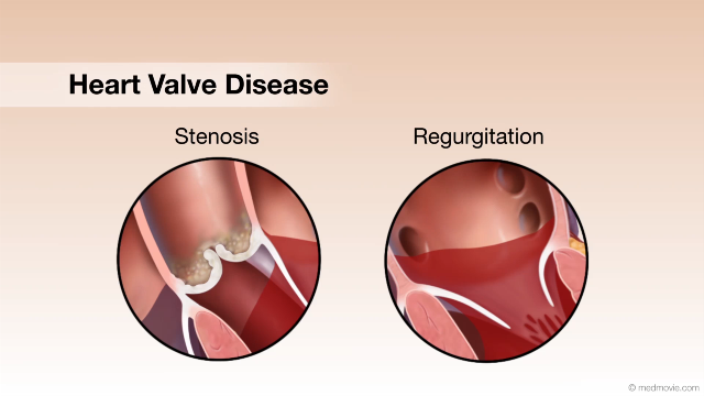 Heart Valve DiseaseValvular heart disease is any condition that disrupts the proper function of the valve. There are four valves in the…
 Heart Valve SurgeryHeart valve replacement surgery replaces an abnormal or diseased heart valve with a healthy one.
There are four…
 Heart ValvesThe heart has four valves. The mitral valve, the tricuspid valve, the pulmonary valve and the aortic valve. The mitral…
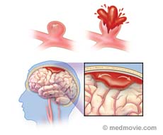 Hemorrhagic StrokeHemorrhagic stroke occurs when an artery in the brain bursts. This blood loss damages the brain. It may also cause a…
 High Blood PressureHigh blood pressure, or hypertension, is a condition in which blood pressure levels are measured above the normal…
 High Blood Pressure ComplicationsBlood pressure is a measurement that reflects the pressure within the arteries. When the heart pumps, the higher…
 High CholesterolCholesterol is a fatty type substance produced by the body and absorbed from the food you eat. Some cholesterol is…
 Holter MonitorA Holter monitor is a battery-operated, portable device that measures and tape-records your heart’s electrical activity…
 Hypertrophic CardiomyopathyThe normal heart has strong muscular walls that pump blood out of the heart.
Hypertrophic cardiomyopathy is a…
 Hypoplastic Left Heart SyndromeHypoplastic left heart syndrome is the result of underdevelopment of the left heart structures and includes a tiny, or…
 ICD DeviceAn Implantable Cardioverter Defibrillator, or ICD, is a battery-powered device that keeps track of your heartbeat, and…
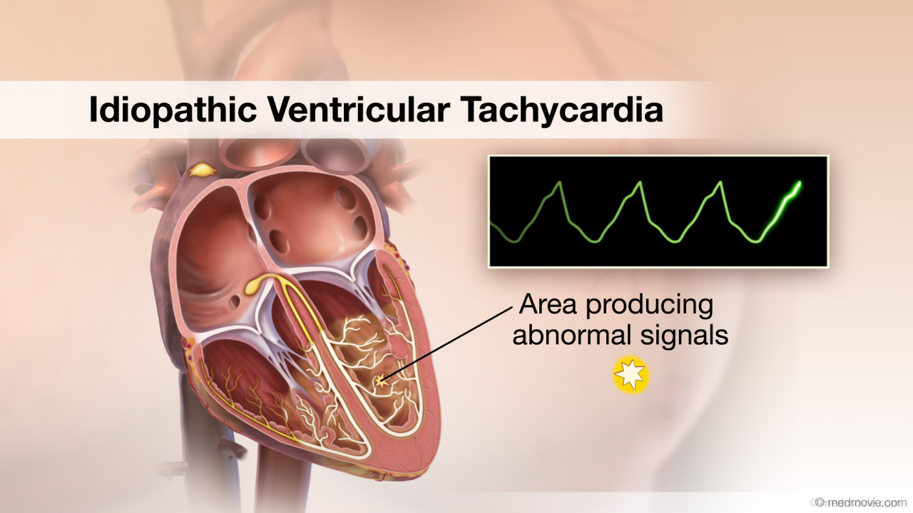 Idiopathic Ventricular TachycardiaThe heartbeat is controlled by the electrical system of the heart. This system is made up of several parts that tell…
 Iliac Artery StentAn iliac stent is a small wire mesh tube that is used to hold open a iliac artery that has been narrowed by artery…
 Insulin ResistanceInsulin resistance is a condition in which the body cannot use insulin efficiently. About 60 million Americans have…
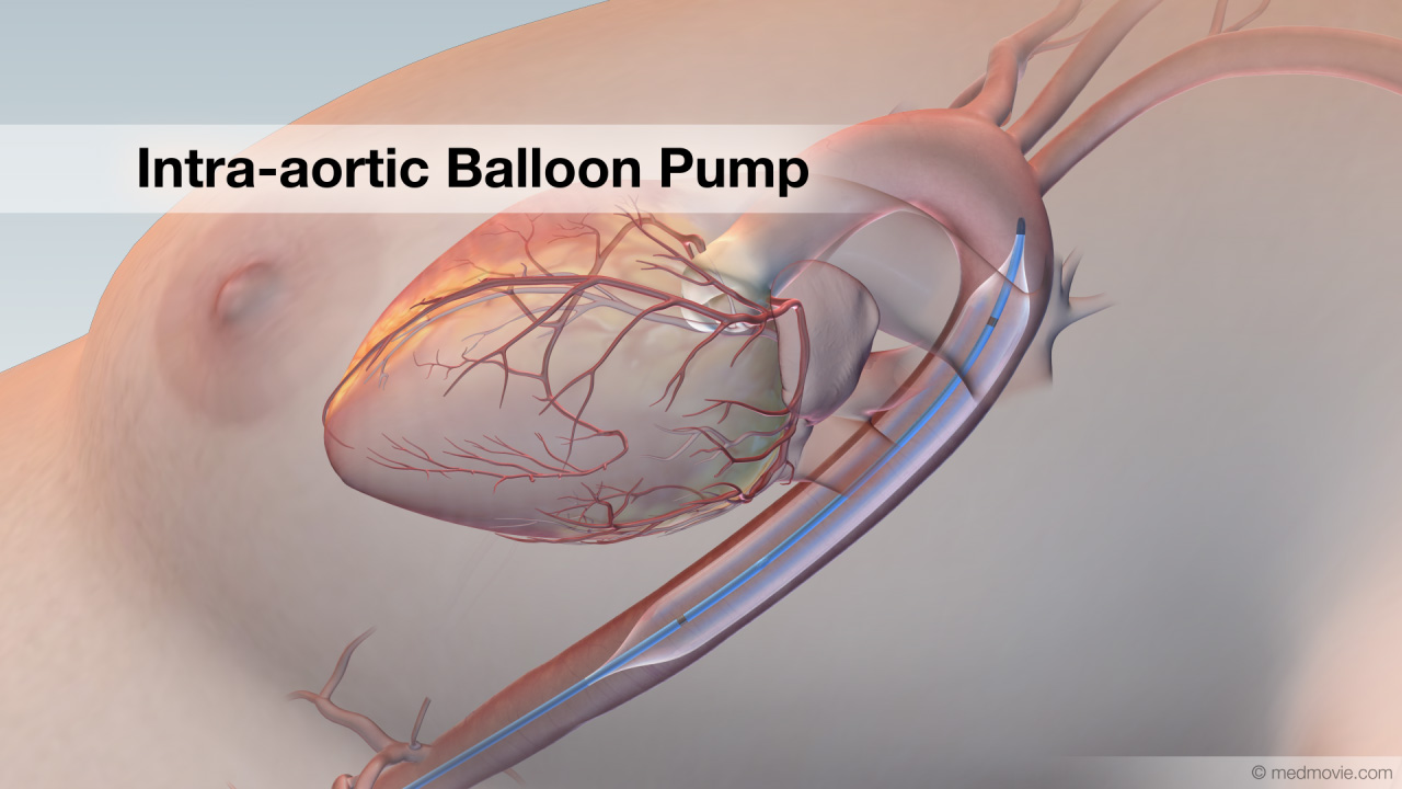 Intra-aortic BalloonAn intra-aortic balloon pump or IABP is an inflatable balloon that is surgically implanted into the aorta to help the…
 Ischemic Ventricular TachycardiaThe heartbeat is controlled by the electrical system of the heart. This system is made up of several parts that tell…
 J Curve PhenomenonWhen the blood pressure or blood cholesterol levels of large groups of people are plotted on a graph against risk of…
 Kawasaki DiseaseKawasaki disease (or syndrome) is a series of symptoms in children that come in phases. Theses symptoms include fever,…
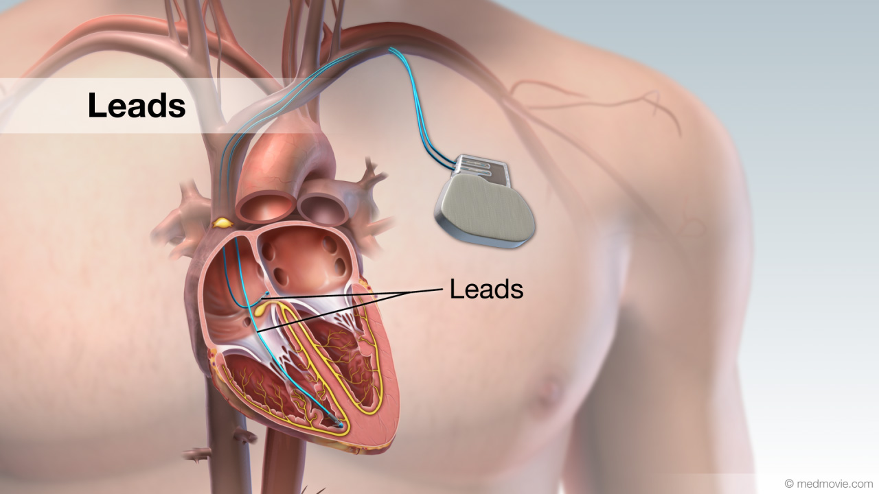 LeadA lead is a device that measures the passage of electrical signals. Leads are used in electrocardiograms to record the…
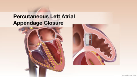 LAACA left atrial appendage closure device can be used for atrial fibrillation patients to help prevent strokes and as an…
 Long QT SyndromeLong QT Syndrome is a condition that is characterized by a delay in repolarization of the heart after the initial…
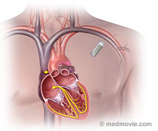 Loop RecorderAn implantable loop recorder is a small device that is implanted under the skin to help identify the causes of…
 Mesenteric Artery StentA mesenteric stent is a small wire mesh tube that is used to hold open a renal artery that has been narrowed by artery…
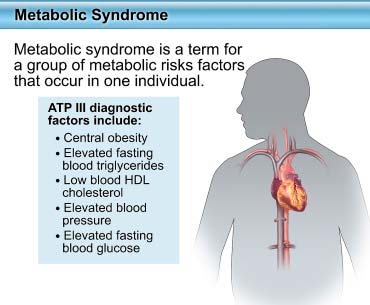 Metabolic SyndromeMetabolic syndrome is a term for a group of metabolic risks factors that occur in one individual. About 35% of US…
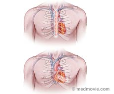 Minimally Invasive Heart SurgeryMinimally invasive heart surgery uses techniques that reduce the size of incisions (cuts) needed to access the heart…
 Mitral ValveThe mitral valve is located between the left atrium and left ventricle of the heart. It allows blood to flow from the…
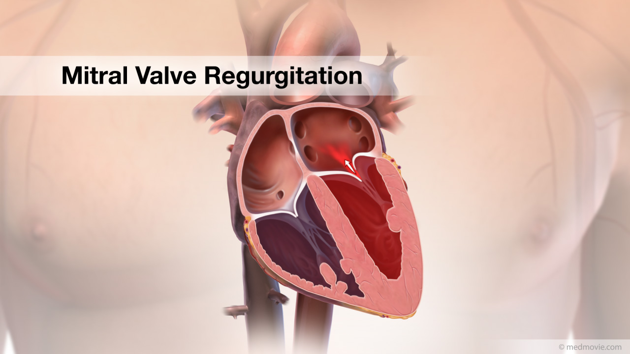 Mitral RegurgitationThe heart has four valves that regulate blood flow through the heart. The mitral valve is the heart valve located…
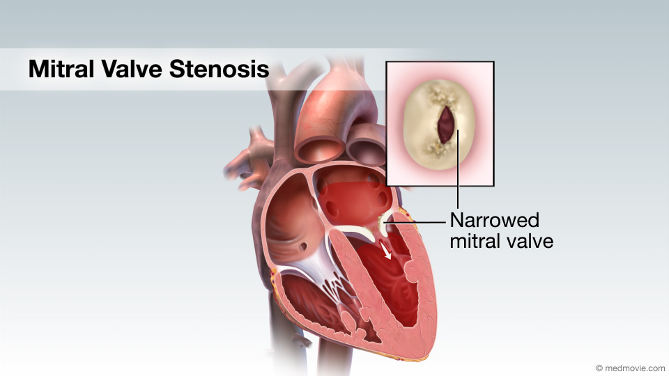 Mitral Valve StenosisMitral valve stenosis is a condition when the mitral valve does not open properly, narrowing the opening, reducing the…
 Blalock-Taussig ModifA Blalock-Taussig shunt is a surgical procedure that helps improve oxygenation of the blood. It is used in cases of…
 MRIMagnetic resonance imaging (MRI) uses a powerful magnetic field, radio waves, and a computer to create detailed…
 Endomyocardial BiopsyEndomyocardial Biopsy — In this test a small amount of tissue is removed from the internal lining of the heart for…
 MPIMyocardial perfusion imaging (MPI) is a diagnostic test used to detect coronary artery disease (blockages). The test…
 Vascular UltrasoundCirculation (Vascular) Ultrasound provides your doctor with moving images of your circulatory system (arteries and…
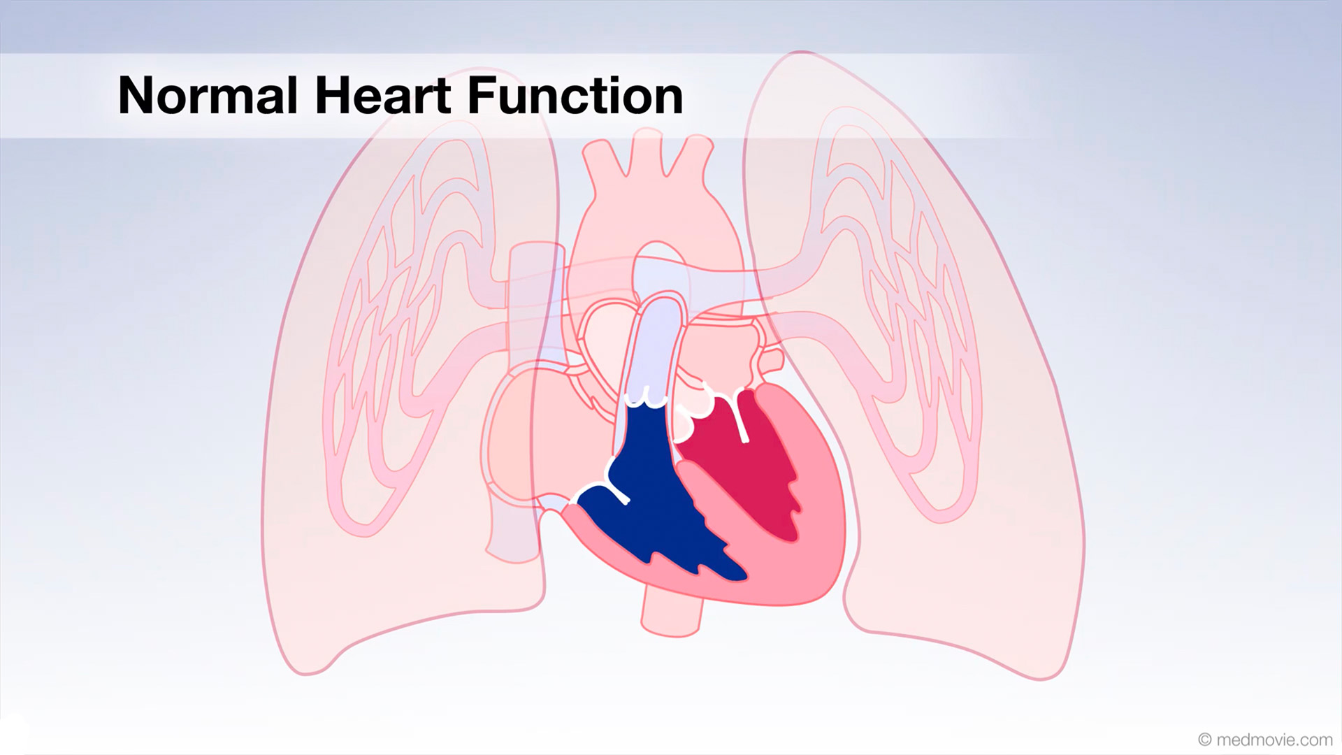 Normal Heart FunctionBlood flows through the heart in a specific pattern in order to pump deoxygenated, or blue blood to the lungs and…
 Norwood ProcedureA Norwood procedure is used to correct a type of congenital heart defect called hypoplastic left heart syndrome. This…
 Open Heart SurgeryOpen-heart surgery is a general term for a surgical procedure that involves making a midline incision in the sternum…
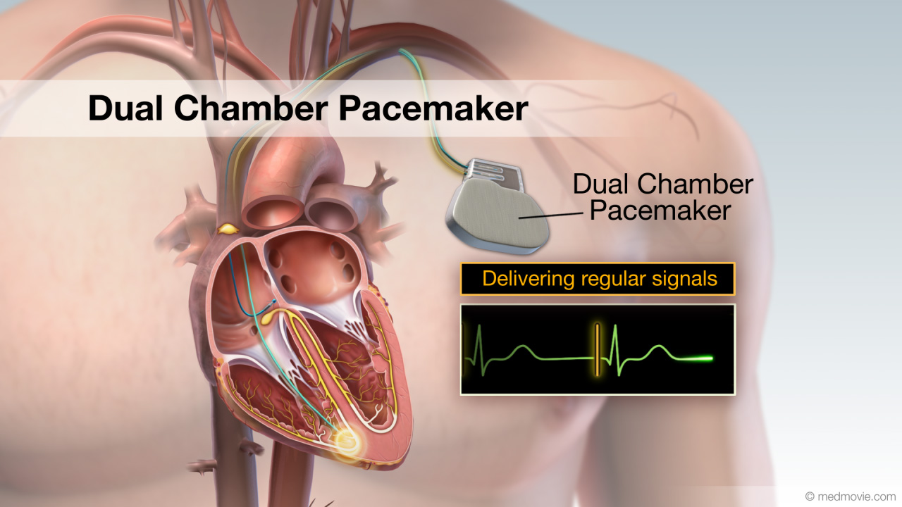 Pacemaker - Dual ChamberA pacemaker is a battery-powered device that sends electrical signals to your heart to help it beat at a proper rate or…
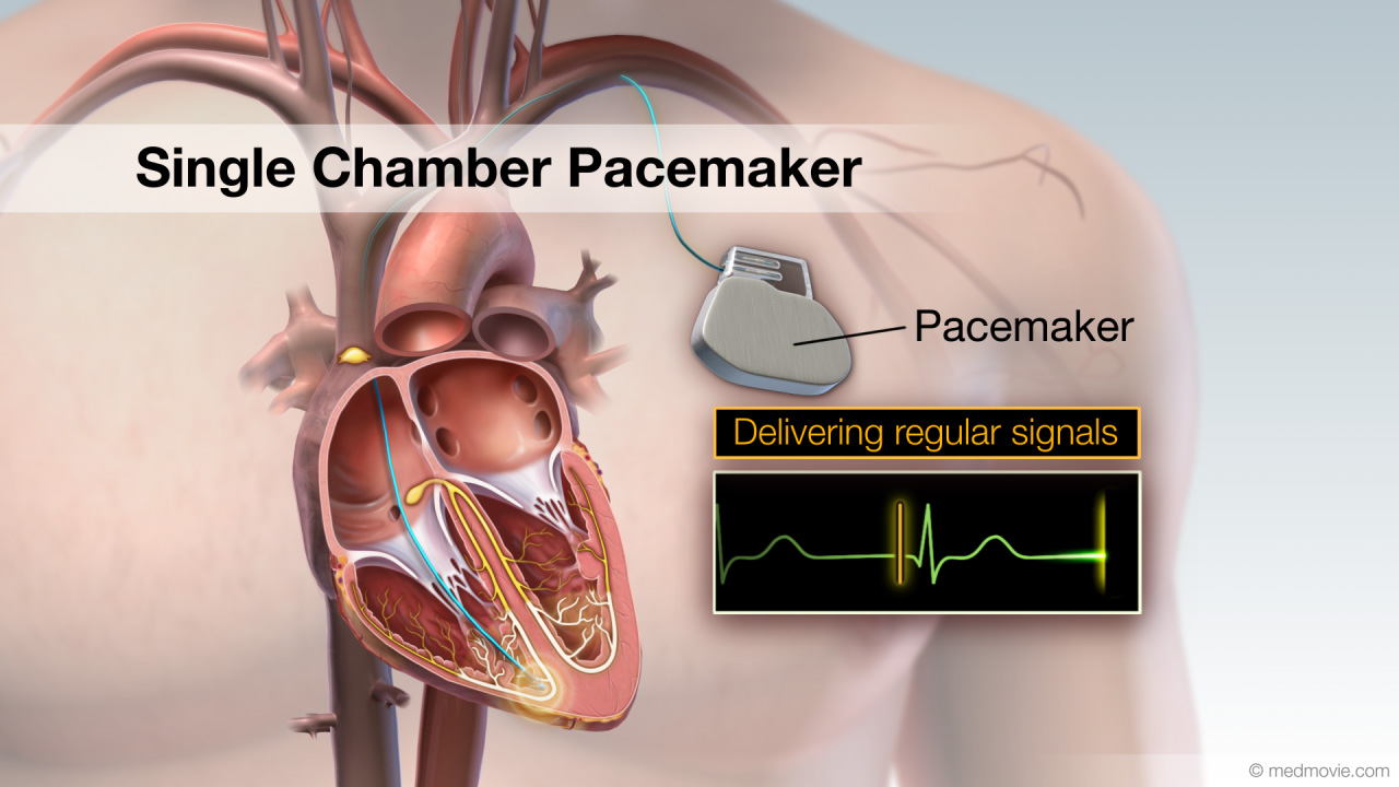 Pacemaker - Single ChamberA pacemaker is a battery-powered device that sends electrical signals to your heart to help it beat at a proper rate or…
 Passive FixationA passive fixation lead is a type of pacemaker or defibrillator “wire” that is used to send the electrical signal of…
 Patent Ductus Arter.The ductus arteriosus is a blood vessel that connects the blood vessel going to the lungs, known as the pulmonary…
 Patent Foramen OvalePatent Foramen Ovale, or PFO, is a hole in the atrial septum that separates the atria. This allows oxygen-poor blood to…
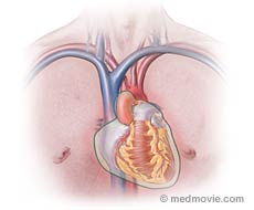 PericarditisPericarditis is an inflammation of the pericardium. The pericardium is the thin layer of tissue that surrounds the…
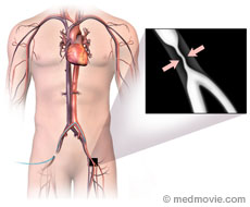 Peripheral AngiogramA peripheral angiogram is a test used to detect blockages in the arteries of the body other than the heart (coronary…
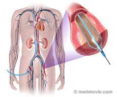 Peripheral AngioplastyPeripheral angioplasty is a procedure that is used to treat narrowed or blocked arteries in the circulation of the…
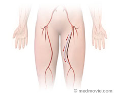 Peripheral Artery BypassA peripheral artery bypass is a procedure that uses a graft to reroute blood around a blockage in a peripheral artery.…
 PADPeripheral arterial disease (PAD) describes any atherosclerotic narrowing of the arteries in the body other than those…
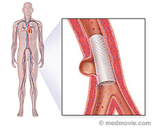 PV Aneurysm Stent ExclusionPeripheral vascular aneurysm stent exclusion is a minimally invasive procedure that is used seal off an aneurysm in an…
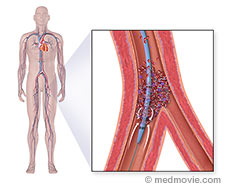 PV Angiojet ThrombectomyPeripheral vasculature angiojet thrombectomy is a minimally invasive procedure that is used to remove blood clots that…
 PV CTO TherapyPeripheral vasculature chronic total occlusion therapy refers to one of several procedures that can be used to treat…
 PV Excisional AtherectomyPeripheral vascular excisional atherectomy is a minimally invasive procedure that is used to remove plaque that has…
 PET ScanPositron Emission Tomography (PET) is a noninvasive nuclear imaging test that uses radioactive tracers to produce…
 Physical ActivityA sedentary lifestyle is one of the major risk factors for cardiovascular disease. A sedentary lifestyle is…
 Plural EffusionPleural effusion is a condition in which fluid builds up in the pleural cavity around the lungs. The lungs are…
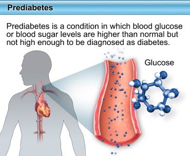 PrediabetesPrediabetes is a condition in which blood glucose (blood sugar) levels are higher than normal but not high enough to be…
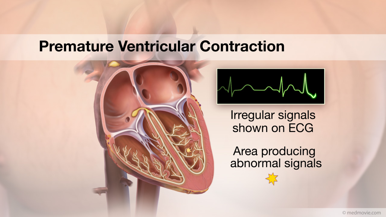 PVCThe heartbeat is controlled by the electrical system of the heart. This system is made up of several parts that tell…
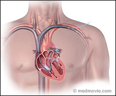 Pulmonary Artery CatheterPulmonary Artery Catheterization (Right Heart Catheterization) is used to obtain diagnostic information about the…
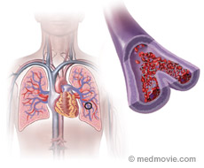 Pulmonary EmbolismA pulmonary embolism is a blood clot that had “traveled” to the lungs, most commonly from blood clots in the legs. The…
 Pulm. Valve StenosisThe pulmonary valve is located between the right ventricle and the large blood vessel to the lungs which is called the…
 Pulmonary VeinsThere are four pulmonary veins that return oxygenated blood from the lungs to the left side of the heart. After blood…
 Quitting SmokingQuitting smoking is not easy, but there are things you can do to increase your chances of success. Planning strategies…
 Radial Cardiac CatheterCardiac catheterization, commonly called a “cardiac cath”, is a procedure used to evaluate multiple aspects of the…
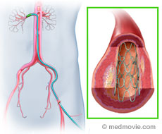 Renal Artery StentA renal stent is a small wire mesh tube that is used to hold open a renal artery that has been narrowed by artery…
 RestenosisRestenosis is a narrowing of an artery following an angioplasty or stenting procedure that was performed to open the…
 Right AtriumThe heart is divided into four chambers. The two upper chambers are the atria, and are separated into the right and…
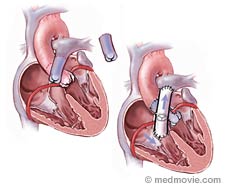 Ross ProcedureA Ross Procedure is surgery of the aortic valve that replaces a diseased or malformed aortic valve with the patient’s…
 RVOT Ventricular TachycardiaThe heartbeat is controlled by the electrical system of the heart. This system is made up of several parts that tell…
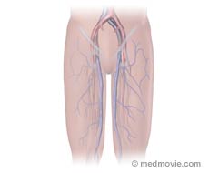 Saphenous VeinThe saphenous vein is the long vein that drains blood from the legs, running from the ankle and up the leg on the inner…
 Sick Sinus SyndromeSick Sinus Syndrome is a condition in which the heart beats too slowly (less than 60 beats per minute). It may be…
 Single VentricleSingle ventricle is a congenital defect in which one ventricle of the heart is abnormally small (hypoplastic). The…
 Sinus RhythmA normal heartbeat produces a regular, identifiable pattern: the P wave, the QRS complex, and the T wave. These…
 Sinus TachycardiaThe heartbeat is controlled by the electrical system of the heart. This system is made of several parts that tell the…
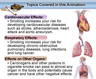 SmokingSmoking tobacco is the leading cause of preventable death in the United States. Smoking has been shown to have harmful…
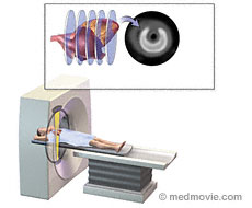 SPECT ScanA SPECT scan of the heart is a noninvasive nuclear imaging test. This test is commonly called a “nuclear stress test”.…
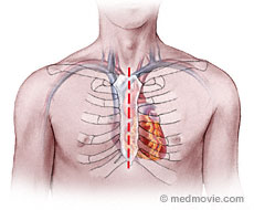 SternotomyA sternotomy is a procedure that is performed to begin open-heart surgery. The sternum (or breastbone) lies vertically…
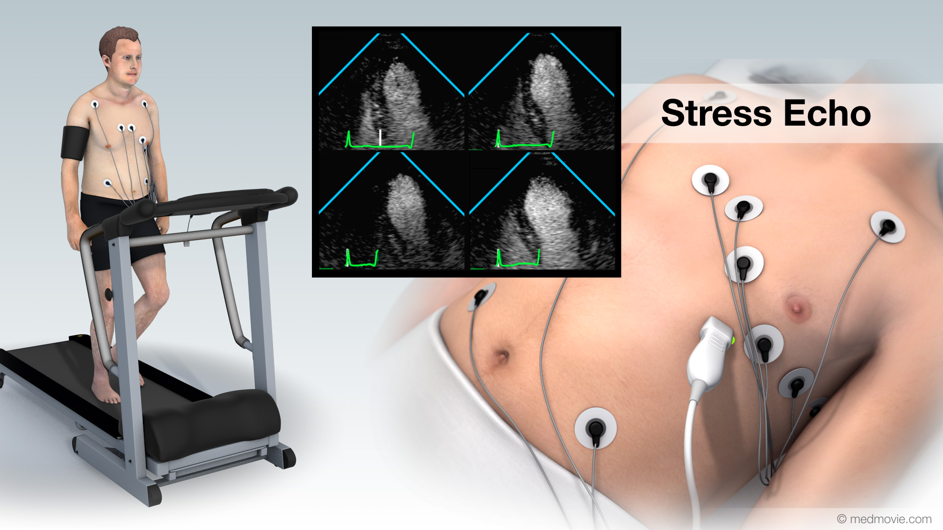 Stress EchoA stress echo, also called an exercise heart ultrasound is used to evaluate heart function by combining an exercise…
 StrokeStroke results from the sudden loss of blood flow to the brain, usually a specific part of the brain (see fig. 1). The…
 Subclavian Artery StentA subclavian artery stent is a small wire mesh tube that is used to hold open a sublcavian artery that has been…
 Sudden Cardiac ArrestSudden cardiac arrest (SCA) is an event caused by a problem with the heart's "electrical" system. SCA occurs when the…
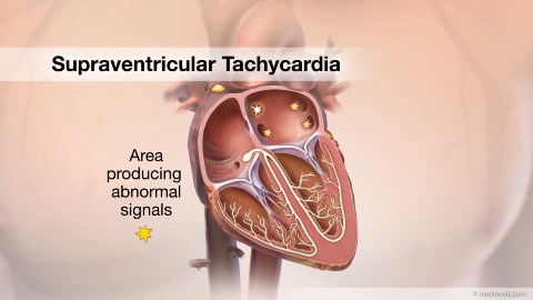 SVTThe heartbeat is controlled by the electrical system of the heart. This system is made up of several parts that tell…
 SyncopeSyncope (or fainting) is a temporary loss of consciousness. It is usually caused by inadequate blood flow to the brain.…
 Tetrology of FallotTetrology of Fallot is characterized by several malformations of the heart. These include: ventricular septal defect…
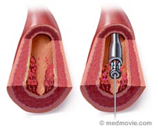 ThrombectomyA thrombectomy is a procedure used to remove a blood clot from a blood vessel. The vessel is usually an artery. A blood…
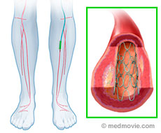 Tibeoperoneal StentA tibeoperoneal stent is a small wire mesh tube that is used to hold open a tibial or peroneal artery that has been…
 TAPVCTotal anomalous venous connections, or TAPVC, is a congenital malformation in which the pulmonary veins join to form a…
 Transcatheter Aortic Valve ReplacementTranscatheter aortic valve replacement, or TAVR, is a treatment for aortic valve stenosis, which is a narrowing of the…
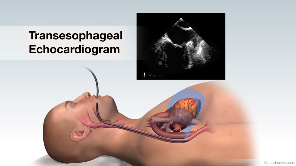 Transesophageal EchocardiogramA transesophageal echocardiogram is a diagnostic test used to view the structures of the beating heart. In this…
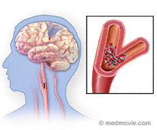 TIAA transient ischemic attack (TIA) is a stroke in which the symptoms resolve quickly, usually spontaneously and usually…
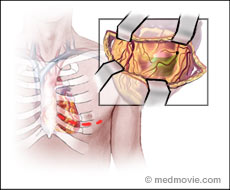 TMRTransmyocardial Revascularization (TMR) is a procedure used to relieve severe angina or chest pain in very ill patients…
 Transp. of Great Arter.D-Transposition (shown in this animation) of the great arteries (TGA) is a congenital heart defect in which the aorta,…
 Tricuspid AtresiaTricuspid atresia with is a congenital defect in which the tricuspid valve that lies between the right atrium of the…
 Tricuspid ValveThe heart has four valves that regulate blood flow through the heart. The mitral valve and the tricuspid valve…
 Truncus ArteriosisTruncus arteriosus is a congenital defect in which the two major arteries (the aorta and the pulmonary artery) fail to…
 Valves- 2 ViewsThe heart has four valves that regulate blood flow through the heart. The mitral valve and the tricuspid valve…
 ValvuloplastyA valvuloplasty is a procedure that is performed to open a narrowed (stenotic) valve. It can be performed using a…
 Vascular ProblemsThe vascular (circulatory) system is made up of blood vessels called arteries and veins. Arteries carry oxygenated…
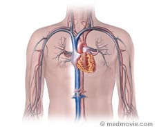 Vena CavaThe vena cava are the largest veins in the body. Veins carry deoxygenated blood back toward the heart. The vena cava…
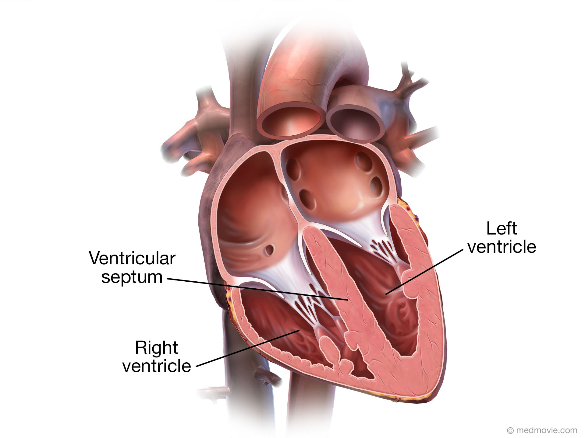 VentriclesThe heart has two sides (a right and a left) and is comprised of four chambers. The two atria, right and left, receive…
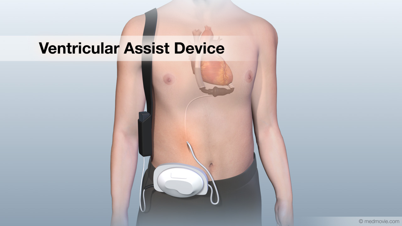 Ventricular Assist DeviceA ventricular assist device, VAD, is a pumping device that is used to help a heart that can no longer pump blood…
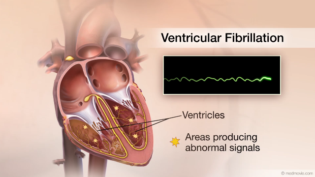 Ventricular FibrillationThe heartbeat is controlled by the electrical system of the heart. This system is made up of several parts that tell…
 Ventric. Septal DefectVentricular septal defect, or VSD, is a hole in the septum that separates the right and left ventricles of the heart.…
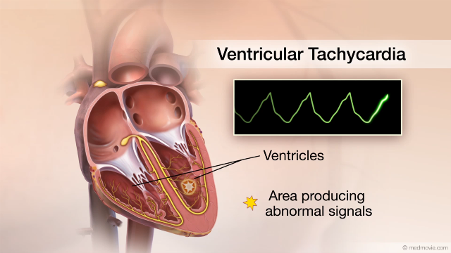 Ventric. TachycardiaThe heartbeat is controlled by the electrical system of the heart. This system is made of several parts that tell the…
 Ventric. Tachycardia AblationVentricular tachycardia ablation is a procedure used to correct ventricular tachycardia by destroying the tissue that…
 WPW SyndromeWolff-Parkinson White Syndrome (WPW) is a condition in which the heart beats too fast due to abnormal, extra electrical…