Myocardial Perfusion Imaging
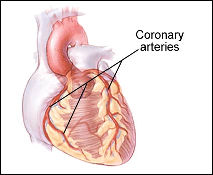
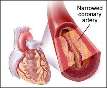
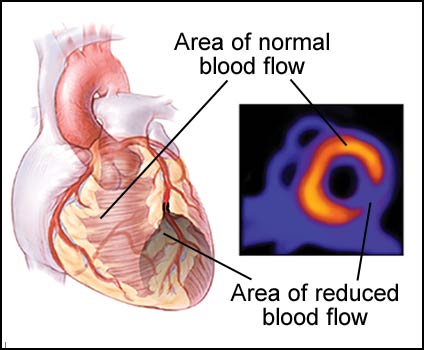
Myocardial perfusion imaging (MPI) is a diagnostic test used to detect coronary artery disease (blockages) (see fig. 1 and 2). The test utilizes a small, safe amount of a radioactive material that is injected into the blood stream during a stress test. This is absorbed by the heart muscle with high amounts absorbed by heart muscle supplied by normal arteries and low amounts absorbed by heart muscle supplied by blocked arteries. This difference between normal and abnormal amounts is detected by a special camera and indicates where a blockage is likely. Usually, the same type of pictures are obtained “at rest”. The differences between the “stress” pictures and “rest” pictures can also indicate if the blockage is bad enough to have caused a heart attack (see fig. 3).
Figure 1: Coronary arteries that supply blood to the heart.
Figure 2: Coronary artery narrowed by atherosclerotic plaque.
Figure 3: Areas of normal and abnormal blood flow revealed by myocardial perfusion imaging. The myocardial image on the right shows a cross-section of the heart muscle
Visit Ohio Heart and Vascular Center for more information.