Atrial Septal Defect
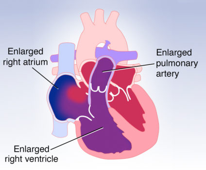
An atrial septal defect, or ASD, is a hole in the wall that separates the upper two chambers of the heart, known as the right and left atria. An ASD allows oxygen-rich blood to flow from the left atrium to right atrium, increasing the volume of blood that flows to the right side of the heart and lungs. In a large ASD, the right atrium, right ventricle, and pulmonary artery become enlarged, or dilated, as a result of the extra blood flow.
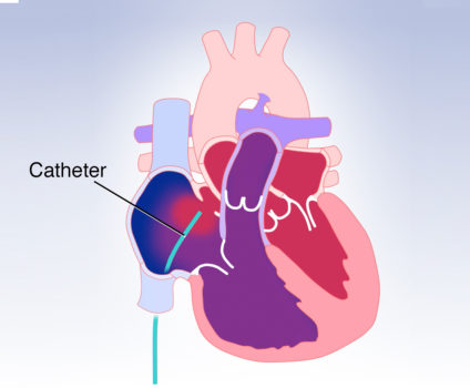
ASDs can be located in different parts of the atrial wall. The most common ASD is located in the secundum atrial septum and can often be closed during a cardiac catheterization. In this procedure, a specially designed catheter is inserted into the large vein in the groin, passed through the inferior vena cava into the right atrium and then into the left atrium. A plug, or device, is then passed through the catheter and expanded within the hole to close it and block blood flow from the left to the right atrium.
Secundum ASDs that are very large or ASDs that are located in other parts of the atrial wall, such as the sinus venosus, or primum atrial septum, are more complex and cannot be closed using a catheter. These holes can be surgically repaired.
Visit Michigan Heart Group for more information.