Peripheral Angiogram
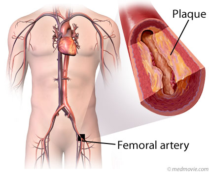
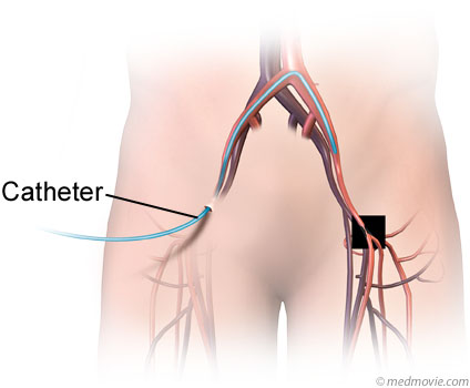
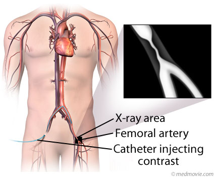
A peripheral angiogram is a test used to detect blockages in the arteries of the body other than the heart (coronary angiogram) and brain (cerebral angiogram). A common reason for performing this test is for suspected blockage in the arteries of the leg (see fig. 1). The test is done by injecting X-ray contrast (dye) into the blood vessels. This allows X-ray pictures to be taken of the blood vessel lumen (the inside of the blood vessel where the blood flows). The contrast is injected through a catheter; a long, thin, hollow tube that is inserted into the circulation through a needle puncture and guided to a position that allows the contrast to flow through the suspected blockage (see fig. 2 through 4) Using this technique, taking pictures of almost any blood vessel of the body is possible.
Figure 1: Peripheral artery disease with narrowing caused by plaque in the femoral artery.
Figure 2: Catheter being guided into the femoral artery.
Figure 3: Angiogram being performed with contrast dye.
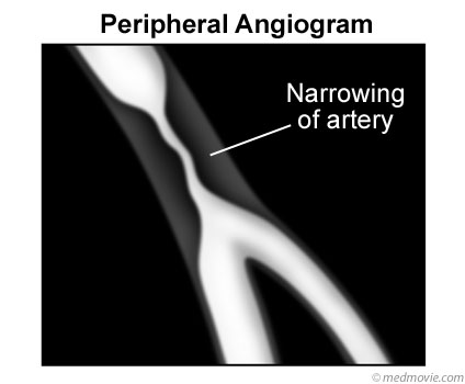
Figure 4: Angiogram with narrowed artery revealed.
Visit Michigan Heart Group for more information.