Tetrology of Fallot
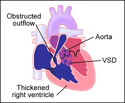
Tetrology of Fallot is characterized by several malformations of the heart. These include: ventricular septal defect (VSD), right ventricular outflow obstruction, resultant right ventricular hypertrophy, and an aorta that lies above the VSD (see fig. 2). The VSD is a hole in the wall that separates the ventricles allowing blood from both chambers to mix. The right ventricular outflow tract is obstructed by a small pulmonary valve combination and a muscular narrowing below the valve. The thickened wall (hypertrophy) due to increased pressure of the right ventricle. The aorta lies above the VSD and carries blood from both chambers to the body. The amount of blood that reaches the lung determines the degree of cyanosis (blueness of the skin).
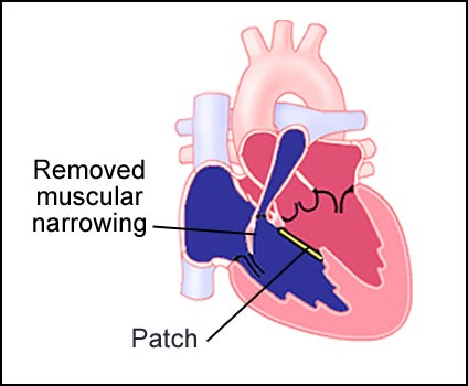
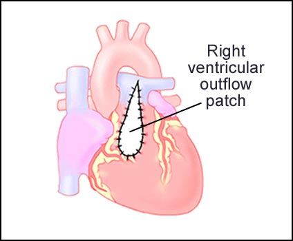
This condition can be surgically repaired. First, the outflow obstruction is removed by cutting away the muscular narrowing. The VSD is repaired with a patch (see fig. 3). When necessary another patch is then placed over the right ventricular outflow tract to widen the opening (see fig. 4). Normal blood flow is restored.
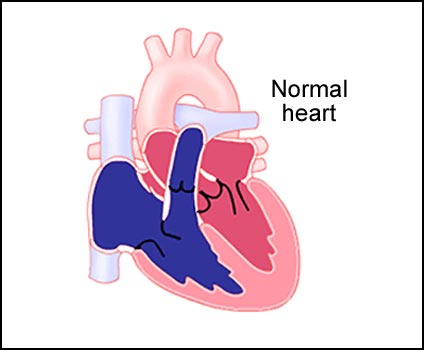
Figure 1: Cut away view of the normal heart.
Figure 2: Heart with Tetrology of Fallot.
Figure 3: First steps of surgical repair. Removal of muscular narrowing, patch to repair of VSD.
Figure 4: Finished surgical repair with right ventricular outflow patch.
Visit Medmovie.com Website for more information.