Transesophageal Echocardiogram
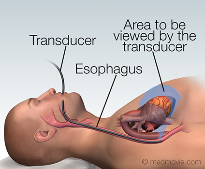
A transesophageal echocardiogram is a diagnostic test used to view the structures of the beating heart. In this procedure, a transducer is passed through the mouth and into the esophagus, which lies behind the heart. The transducer then sends images of the heart to a monitor. This imaging process is called an echocardiogram. An echocardiogram uses ultrasound waves to create moving pictures of the structures of the heart such as the chambers and valves. It can also evaluate blood flow and pressures within the heart with a special technique called Doppler Echocardiography.
Figure 1: Transducer in place inside the esophagus.
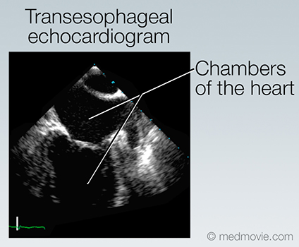
Figure 2: Images from the transducer are sent to a monitor.
Figure 3: The images show the moving anatomy of the heart. Abnormalities of the heart may be revealed by these images.
Visit Medmovie.com Website for more information.