SPECT Scan
A SPECT scan of the heart is a noninvasive nuclear imaging test. It uses radioactive tracers that are injected into the blood to produce pictures of the heart. Doctors use SPECT to diagnose coronary artery disease and find out if a heart attack has occurred. SPECT can show how well blood is flowing to the heart and how well the heart is working.
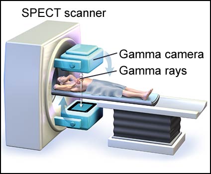
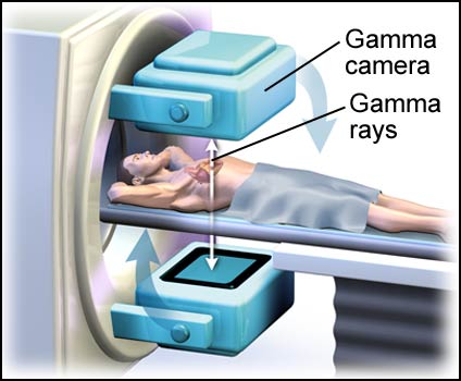
To begin a SPECT scan, radioactive tracers are injected into the patient’s blood. The patient then lies on a table under the camera of the scanner. The tracers are taken up by living heart tissue and produce a type of energy called gamma rays. A gamma camera picks up these rays and a computer converts them into images of the heart (see fig. 1 and 2). These images are produced in “slices” that can be produced from different directions and angles. The images can be used to create a 3-dimensional image. Areas of lightness or darkness or areas of colors represent the amount of tracers taken up by the heart and the relative blood flow reaching that tissue (see fig. 3).
Figure 1: A SPECT scanner taking images of the heart.
Figure 2: The gamma camera recording gamma rays from the heart tissue.
Figure 3: “Sliced” images of the heart that reveal an area of reduced blood flow to the heart.
Visit Medmovie.com Website for more information.