Transposition of the Great Arteries
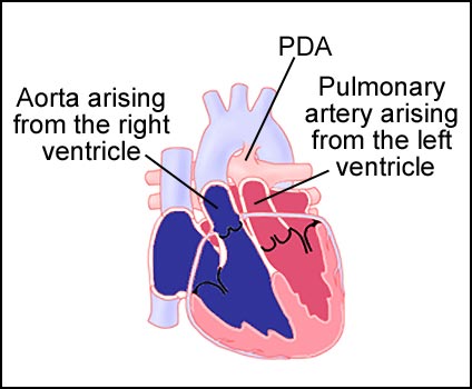
D-Transposition of the great arteries (TGA) is a congenital heart defect in which the aorta, which normally arises from the left ventricle, emerges from the right ventricle instead (see fig. 2). The pulmonary artery, which normally arises from the right ventricle, comes from the left ventricle instead. As a result, oxygen-poor blood is pumped to the body and oxygen-rich blood is pumped to the lungs. Other defects are commonly associated with TGA including: atrial septal defect (ASD), ventricular septal defect (VSD), and/or patent ductus arteriosus (PDA). There must be a mixing site(s) from for survival in the neonatal period. In L- Transposition of the great arteries (corrected transposition), both arteries and ventricles are transposed; thus the circulation is normalized or “corrected”. However, other associated cardiac defects are almost always present.
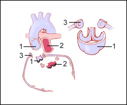
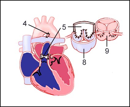
D-Transposition can be surgically corrected by an atrial switch procedure. The coronary arteries are cut away from the base of the aorta. The aorta and pulmonary artery are divided (see fig. 3). The PDA is divided and tied off. The pulmonary artery is then moved in front of the aorta to the right ventricle. The coronary arteries are attached to the neo-aorta that has now become the aortic valve (see fig. 4).
Figure 1: Cut away view of the normal heart.
Figure 2: Transposition of the great arteries.
Figure 3: First steps of surgical repair.
Figure 4: Finished surgical repair.
1. Aorta.
2. Pulmonary artery.
3. Coronary arteries.
4. PDA divided and tied off.
5. Pericardial patch.
6. Pulmonary artery reconstruction.
7. Aortic reconstruction.
Visit Medmovie.com Website for more information.