Truncus Arteriosis
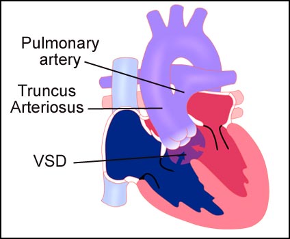
Truncus arteriosus is a congenital defect in which the two major arteries (the aorta and the pulmonary artery) fail to separate leaving one large embryonic vessel that stem from both ventricles. The large artery, known as the truncus arteriosus, has one valve that carries blood from both ventricles. A ventricular septal defect (VSD) is a hole in the wall (septum) between the ventricles that allows blood to flow into the truncus arteriosus.
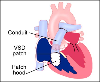
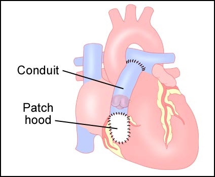
This defect can be surgically repaired. A conduit from the right ventricle to the pulmonary artery in sutured into place. This conduit carries blood from the right ventricle to the pulmonary artery. A patch hood is placed over the conduit origin on the right ventricle. Occasionally, a direct connection between the right ventricle and the pulmonary artery is utilized. The VSD is repaired with a patch. Normal blood flow is then restored.
Figure 1: Cut away view of the normal heart.
Figure 2: Heart with truncus arteriosus.
Figure 3: Finished surgical repair from the interior.
Figure 4: Finished surgical repair.
Visit Medmovie.com Website for more information.