Tricuspid Atresia
Tricuspid atresia with is a congenital defect in which the tricuspid valve that lies between the right atrium of the heart and the right ventricle fails to develop. Blood is able to leave the right atrium through a communication between the atria know as an atrial septal defect (ASD), persistent fetal channel, or patent foramen ovale (PFO). Tricuspid atresia almost always includes a ventricular septal defect (VSD) which allows blood to reach the pulmonary artery. The result of this combination of defects is variable degrees of low oxygen levels in the body.
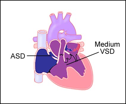
If tricuspid atresia is accompanied by a medium sized VSD, this allows enough blood to flow through the pulmonary artery to the lungs. This size of VSD will keep oxygen levels in safe range, but will not overload the lungs with blood volume (see fig. 3). If tricuspid atresia is accompanied by a large sized VSD, too much blood may flow to the lungs (see fig. 2). If persistant, this would overload the ventricle and damage the pulmonary arteries. When severe cyanosis (blueness of the skin) is present, this condition can be helped in early infancy by using a modified Blalock-Taussig surgical shunt (see fig. 2). Blood from the aorta to the right pulmonary artery improving oxygen levels. If a persistant large VSD is present, a pulmonary band may be placed to limit blood flow (fig. 4).
Figure 1: Cut away view of the normal heart.
Figure 3: Triscuspid atresia with small VSD and Blalock-Taussig shunt.
Figure 2: Triscuspid atresia with medium VSD.
Figure 4: Triscuspid atresia with large VSD and pulmonary artery band.
Visit Medmovie.com Website for more information.