
3D proximal isovelocity surface area
©2026 Medmovie.com. All rights reserved. Medmovie.com creates and licenses medical illustrations and animations for educational use. Our goal is to increase your understanding of medical terminology and help communication between patients, caregiver and healthcare professionals. The content in the Media Library is for your information and education purposes only. The Media Library is not a substitute for professional medical advice, diagnosis or treatment for specific medical conditions.
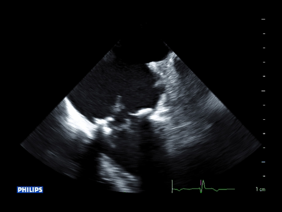 2D TEE 0 degrees MV2D mid-esophageal TEE view of the mitral valve at 0 degrees demonstrating flail prosthetic valve leaflet.
 2D TEE 0 degrees MV with color2D mid-esophageal TEE view of the mitral valve at 0 degrees demonstrating the regurgitant jet associated with the flail…
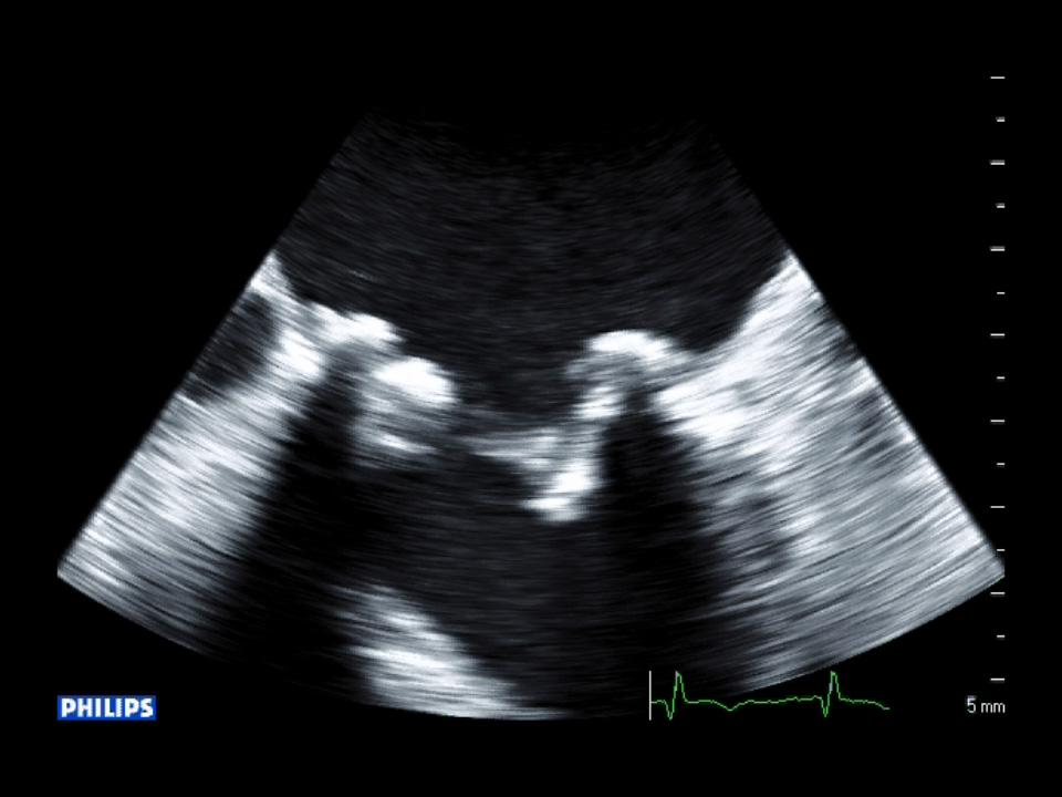 2D TEE 45 degrees MV2D mid-esophageal TEE view of the mitral valve at 45 degrees demonstrating flail prosthetic valve leaflet, vegetation…
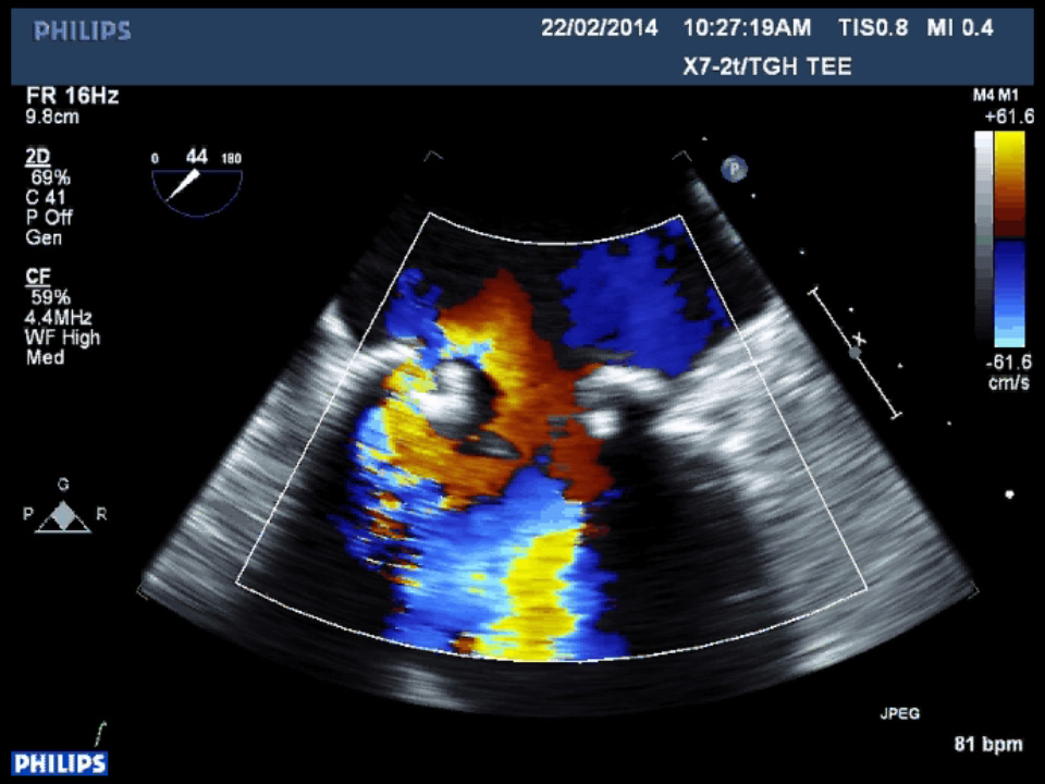 2D TEE 45 degrees MV with color2D mid-esophageal TEE view of the mitral valve at 45 degrees with two regurgitant jets, one is from the flail leaflet…
 2D TEE 75 degrees MV2D mid-esophageal TEE view of the mitral valve at 75 degrees demonstrating that two of the leaflets are flail with…
 2D TEE 75 degrees MV with color2D mid-esophageal TEE view of the mitral valve at 75 degrees demonstrating the presence of a paravalvular leak as flow…
 2D TEE 76 degrees MV2D mid-esophageal TEE view of the mitral valve at 76 degrees demonstrating that a second perforation is present on…
 2D TEE 76 degrees MV with color2D mid-esophageal TEE view of the mitral valve at 75 degrees demonstrating that two of the leaflets are flail with…
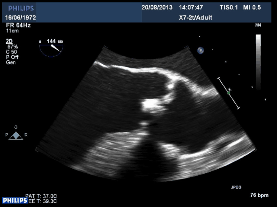 2D TEE at 120 degrees, zoomed on the AV2D mid-esophageal TEE view of the aortic valve at 120 degrees demonstrating thickening and abnormal opening of the…
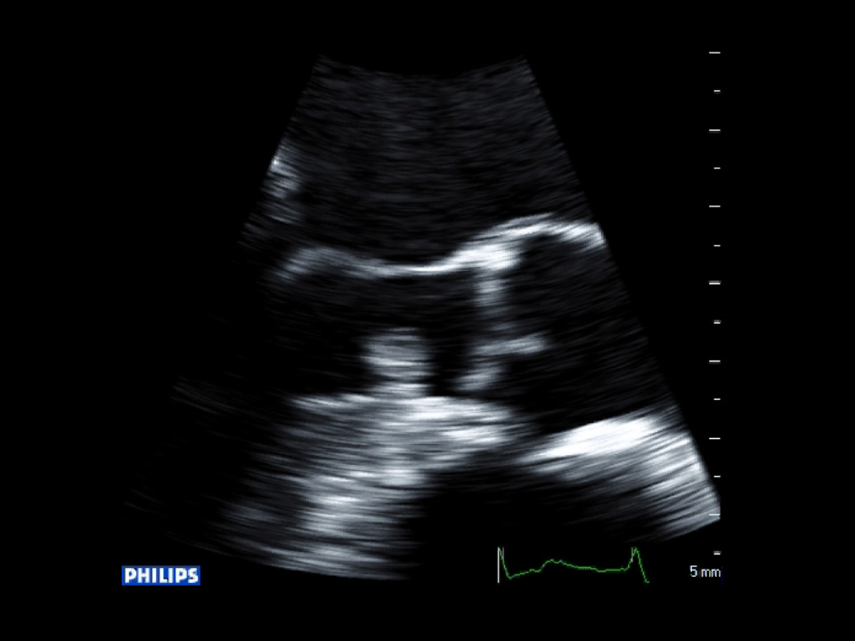 2D TEE at 120 degrees, zoomed on the LVOT2D mid-esophageal TEE view of the left ventricular outflow tract at 120 degrees demonstrating the presence of a…
 2D TEE at 45 degrees, zoomed on the AV2D mid-esophageal TEE view of the aortic valve at 45 degrees demonstrating a single opening.
 2D TEE at 45 degrees, zoomed on the AV with color2D mid-esophageal TEE view of the aortic valve at 45 degrees demonstrating valvular regurgitation.
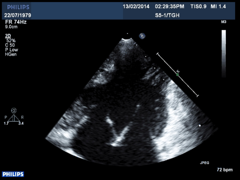 2D TTE apical 4-chamber view2D TTE apical view demonstrating the univentricle with both atrioventricular valve on the same plane.
 2D TTE apical 4-chamber view2D TTE apical 4-chamber view demonstrating systolic anterior motion of the anterior mitral valve leaflet.
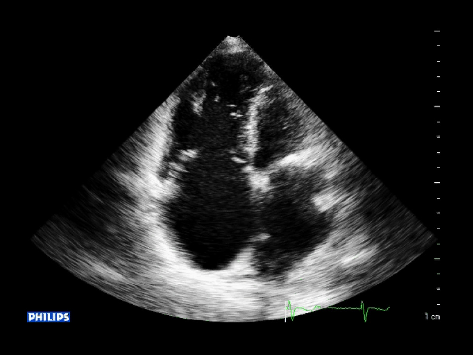 2D TTE apical 4-chamber view of the tricuspid valve2D TTE apical 4-chamber viewof the tricuspid valve demonstrating malcoaptation between the septal and anterior leaflets…
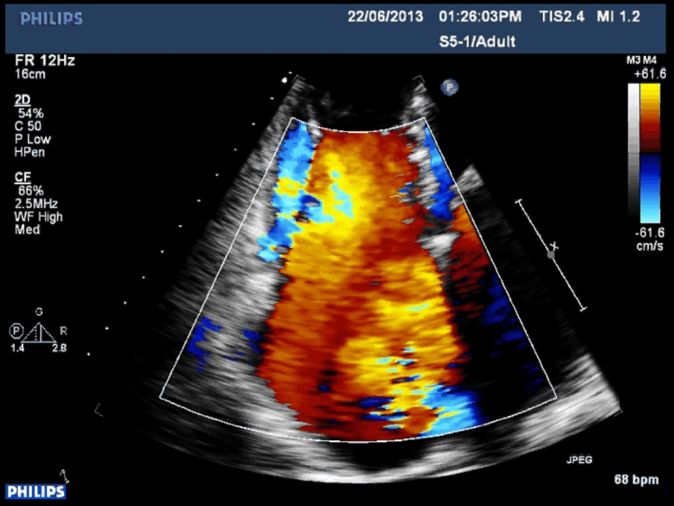 2D TTE apical 4-chamber view with color of the tricuspid valve2D TTE right ventricular inflow view with color demonstrating severe tricuspid regurgitation.
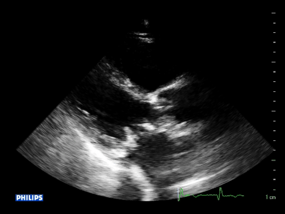 2D TTE PLAX2D TTE parasternal long axis image of the bioprosthetic mitral valve.
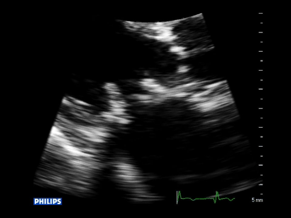 2D TTE PLAX zoom MV2D TTE parasternal long axis view zoomed image of the bioprosthetic mitral valve demonstrating leaflet abnormality.
 2D TTE PLAX zoom MV w/without color2D TTE parasternal long axis view zoomed image of the bioprosthetic mitral valve demonstrating regurgitation through…
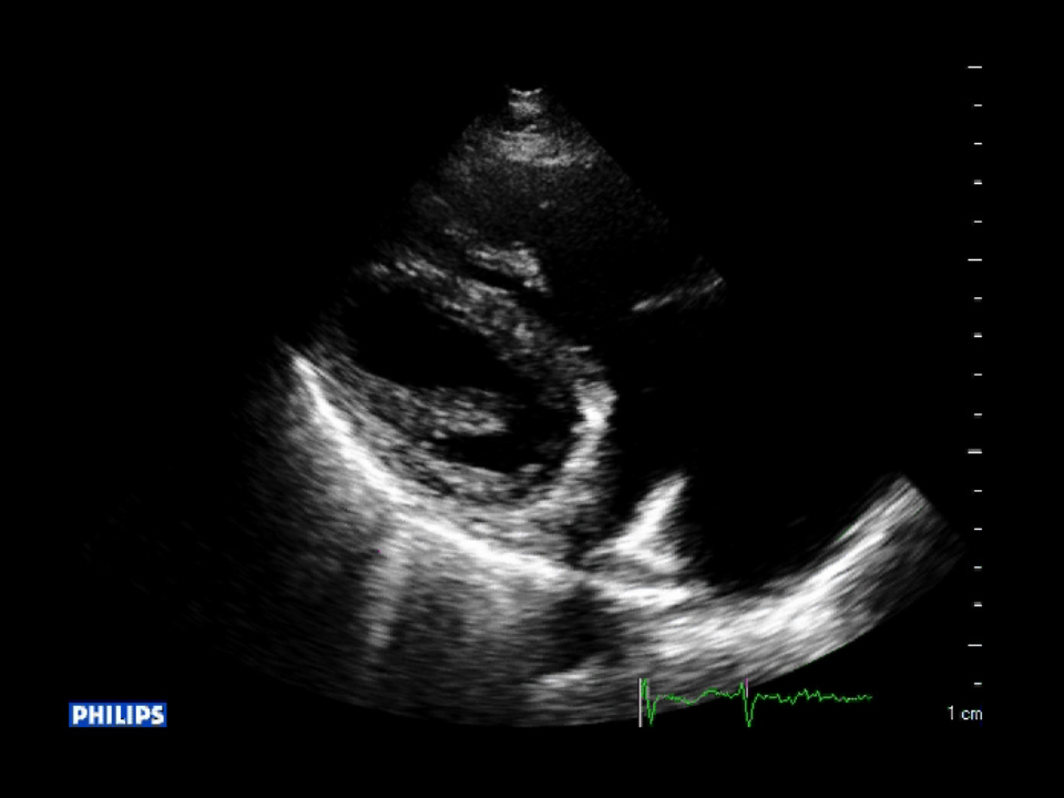 2D TTE RV inflow view2D TTE right ventricular inflow view demonstrating malcoaptation between the septal and anterior leaflets of the…
 2D TTE RV inflow view with color2D TTE right ventricular inflow view with color demonstrating severe tricuspid regurgitation.
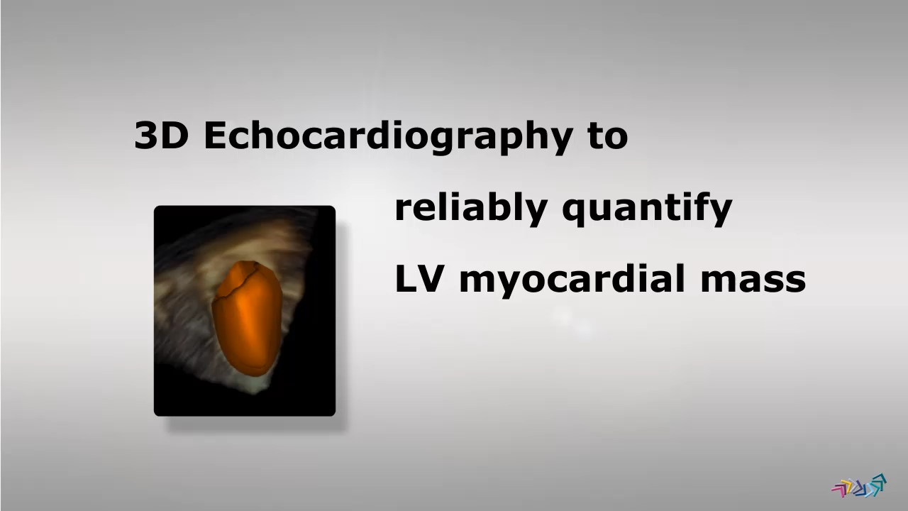
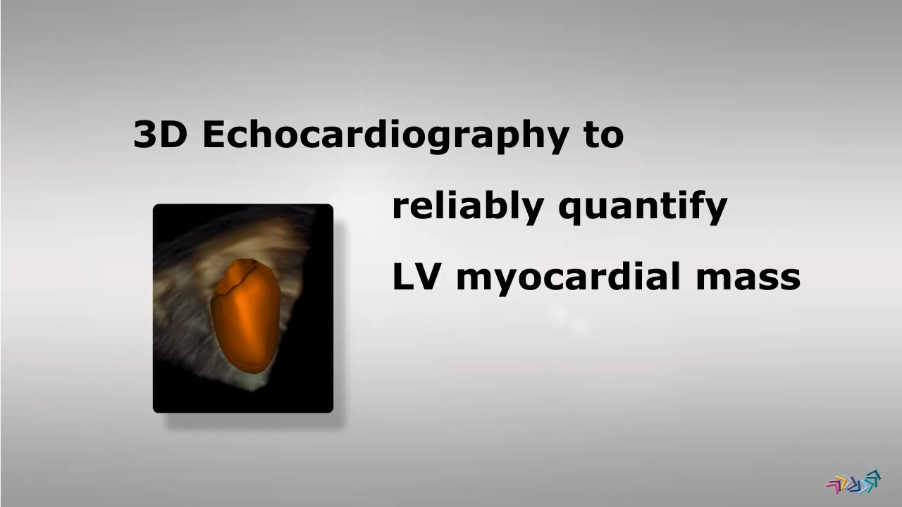 3D Calculation of LV mass3D model of left ventricle for measurement of left ventricular mass
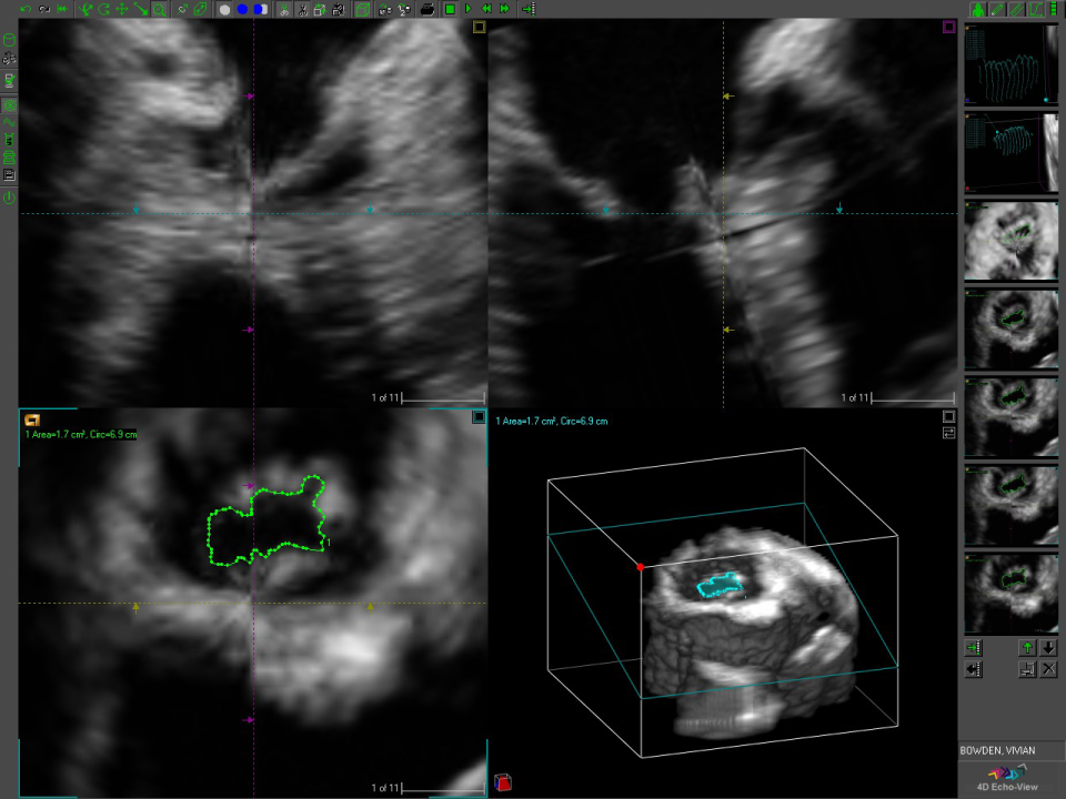 3D evaluation of mitral stenosisUse of 3D echocardiography for measuring anatomic orifice on mitral stenosis
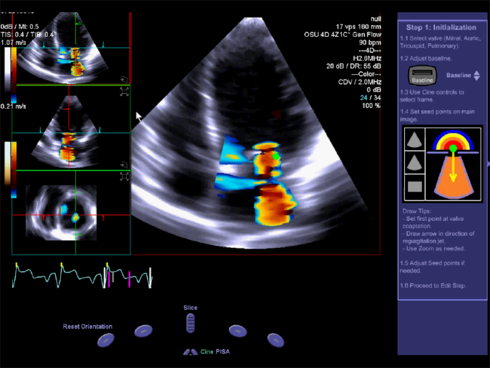 3D Proximal Isovelocity Surface Area3D proximal isovelocity surface area
 3D StrainComprehensive 3D strain analysis
 3D TEE MV en face LA view3D TEE en face view of the mitral valve from the left atrial perspective, demonstrating flail of two leaflets with…
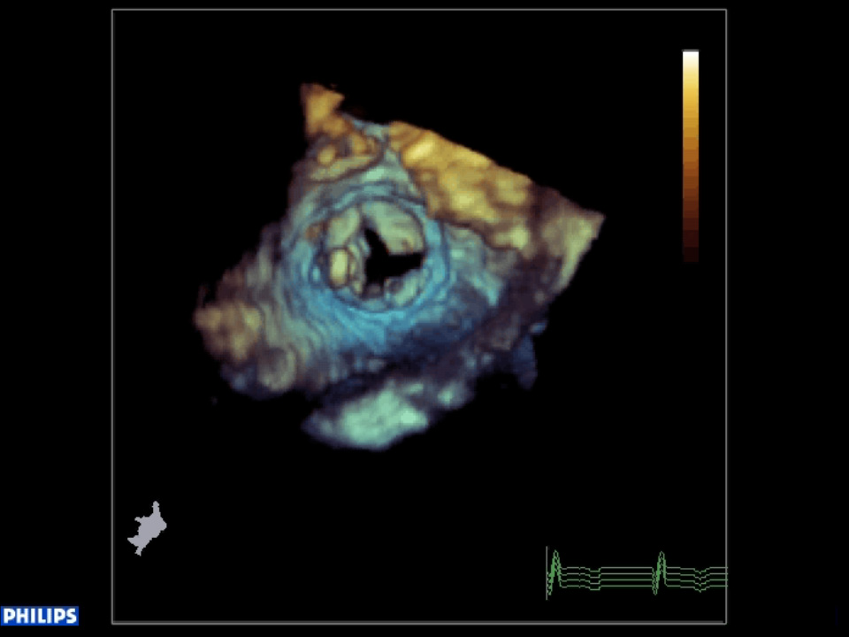 3D TEE MV en face LV view3D TEE en face view of the mitral valve from the left ventricular perspective, demonstrating the infected leaflet's…
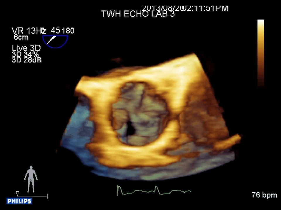 3D TEE view of the AV from the ascending aorta perspective3D TEE view of the aortic valve from the ascending aorta perspective, demonstrating the unicommissural opening and two…
 3D TEE view of the mass from the left ventricular perspective3D TEE view of the mass from the left ventricular perspective, demonstrating its position on the ventricular septum.
 3D TEE view of the mass from the LVOT3D TEE zoomed view of the mass from the left ventricular perspective, demonstrating its position on the ventricular septum.
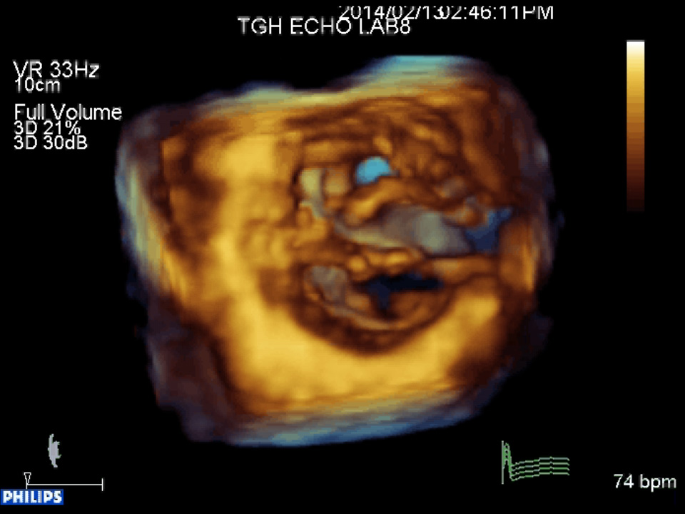 3D TTE view of the atrio-ventricular valves from the ventricular perspective3D TTE view of the atrioventricular valves from the ventricular perspective demonstrating the absence of a ventricular…
 3D TTE view of the mitral valve from the left ventricle3D TTE view of the mitral valve from the left ventricular perspective demonstrating systolic anterior motion of the…
 3D TTE view of the tricuspid valve from the right ventricular perspective3D TTE view of the tricuspid valve from the right ventricular perspective demonstrating that the anterior leaflet moves…
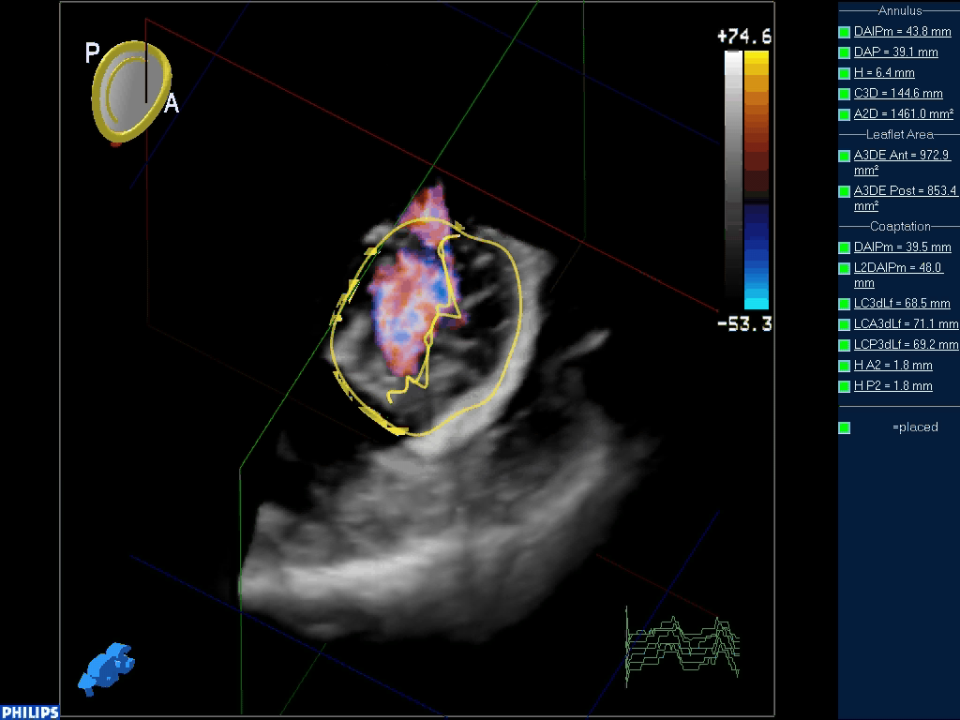 4D image of Mitral regurgitation with multiple jets4D image of mitral regurgitation with multiple jets. Planimetry of proximal jets done to measure orifice areas.
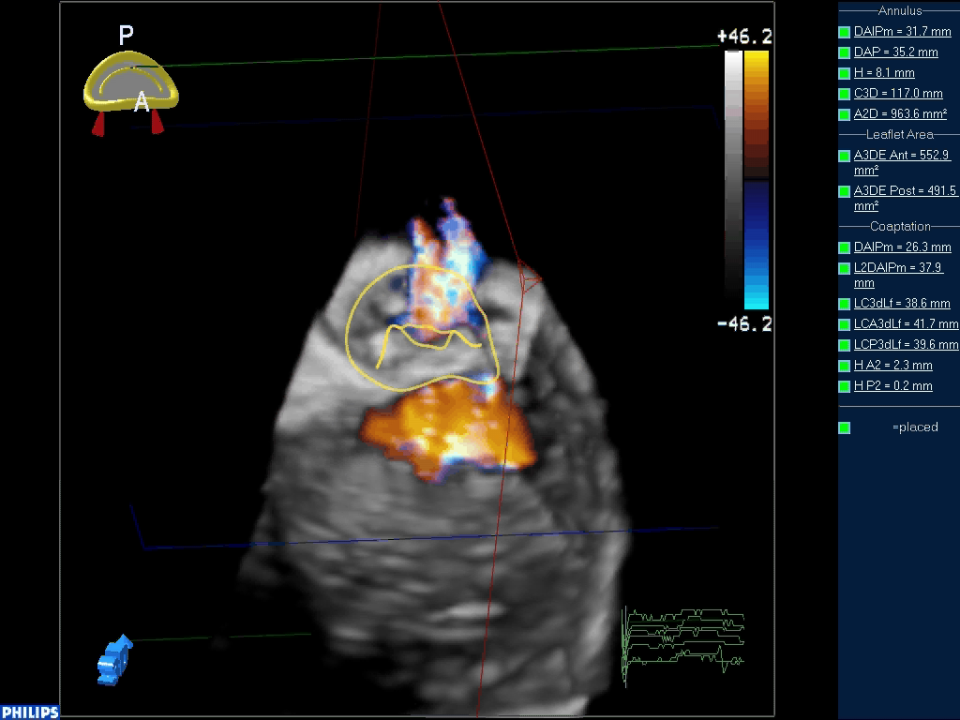 4D image of Mitral regurgitation with single jet4D image of mitral regurgitation with single jet. Planimetry of proximal jets done to measure orifice area.
 4D image of normal mitral valve4D model of mitral valve in systole.
 4D right ventricle evaluation4D evaluation of right ventricle
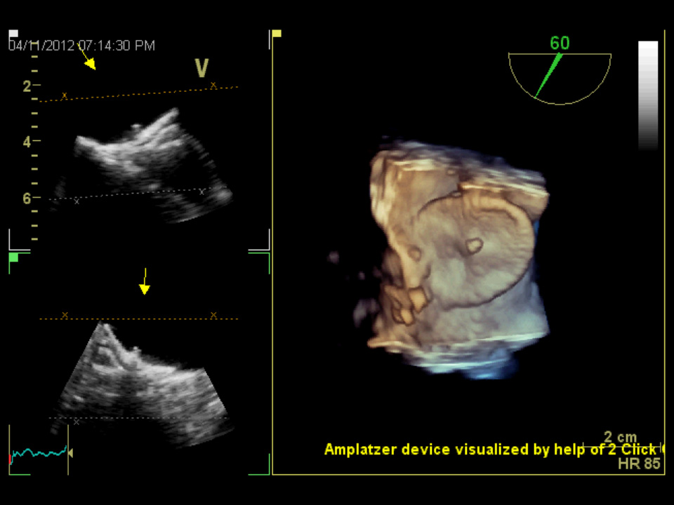 Amplatzer ASD occluder deviceReal time 3D-TEE of an Amplatzer ASD occluder device visualized from the left atrial side. Image acquired using the 2…
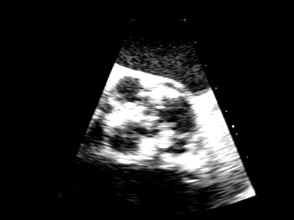 Aortic valve perforation 2DTricuspid valve with thickened leaflets and nodular calcification of the free edges.
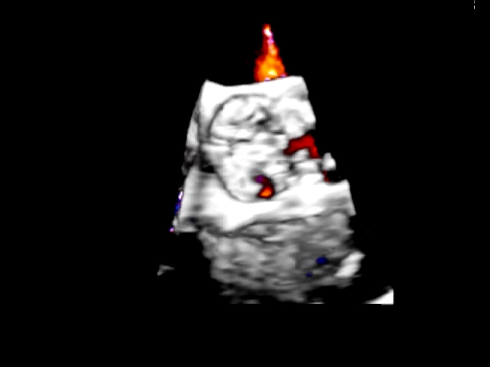 Aortic valve perforations 3DPerforations involving all three cusps. Color flow Doppler confirmation is necessary to rule out dropout artifacts.
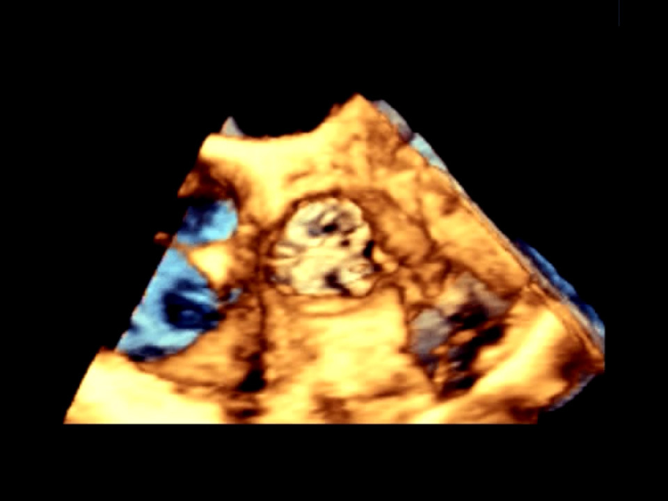 Aortic valve perforations 3D CFDPerforations involving all three cusps with secondary regurgitation.
 Failed mitral valve repair 3DEnface left atrial or surgeon’s view of the mitral valve post repair showing leaflet mal-coaptation in particular…
 Failed mitral valve repair 3D CFDEnface left atrial or surgeon’s view of the mitral valve post mitral repair showing residual mitral regurgitation. …
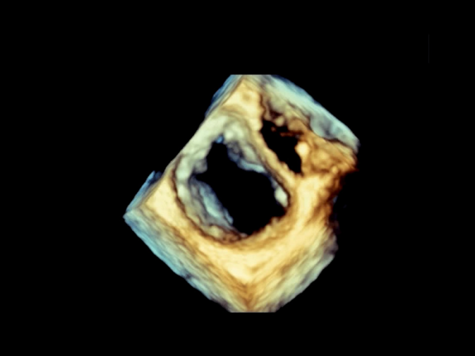 Flail P2 scallop 3DEnface left atrial view of the mitral valve showing a flail P2 segment with two ruptured chords.
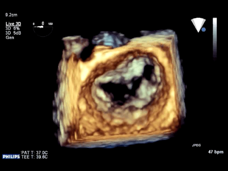 Functional mitral regurgitation 3DEnface left atrial view of the mitral valve showing complete lack of leaflet coaptation.
 Mitral bioprosthesis stenosis 3D LA viewEnface left atrial view of a stenosed mitral bioprosthesis. The aortic valve anteriorly and left atrial appendage…
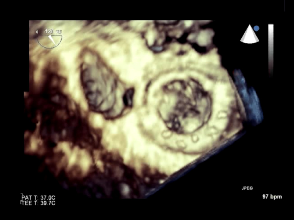 Mitral bioprosthesis stenosis 3D LV viewEnface LV view of a stenosed mitral bioprosthesis showing the three prosthesis struts in the left ventricle.
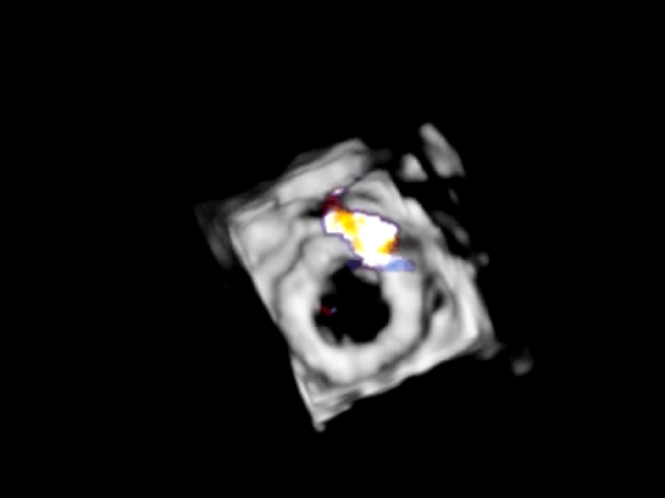 Mitral periprosthetic leak 3D CFDEnface left atrial view of a mitral prosthesis with an anterior periprosthetic leak.
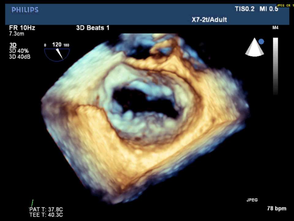 Mitral valve endocarditis on Real time 3D-TEEReal time 3D-TEE of the mitral valve showing a vegetation attached to the P2 leaflet.
 Mitral valve on real time 3D-TEEAppearance of a normal mitral valve on real time 3D-TEE. This shows the “surgeon’s view” of the mitral valve with the…
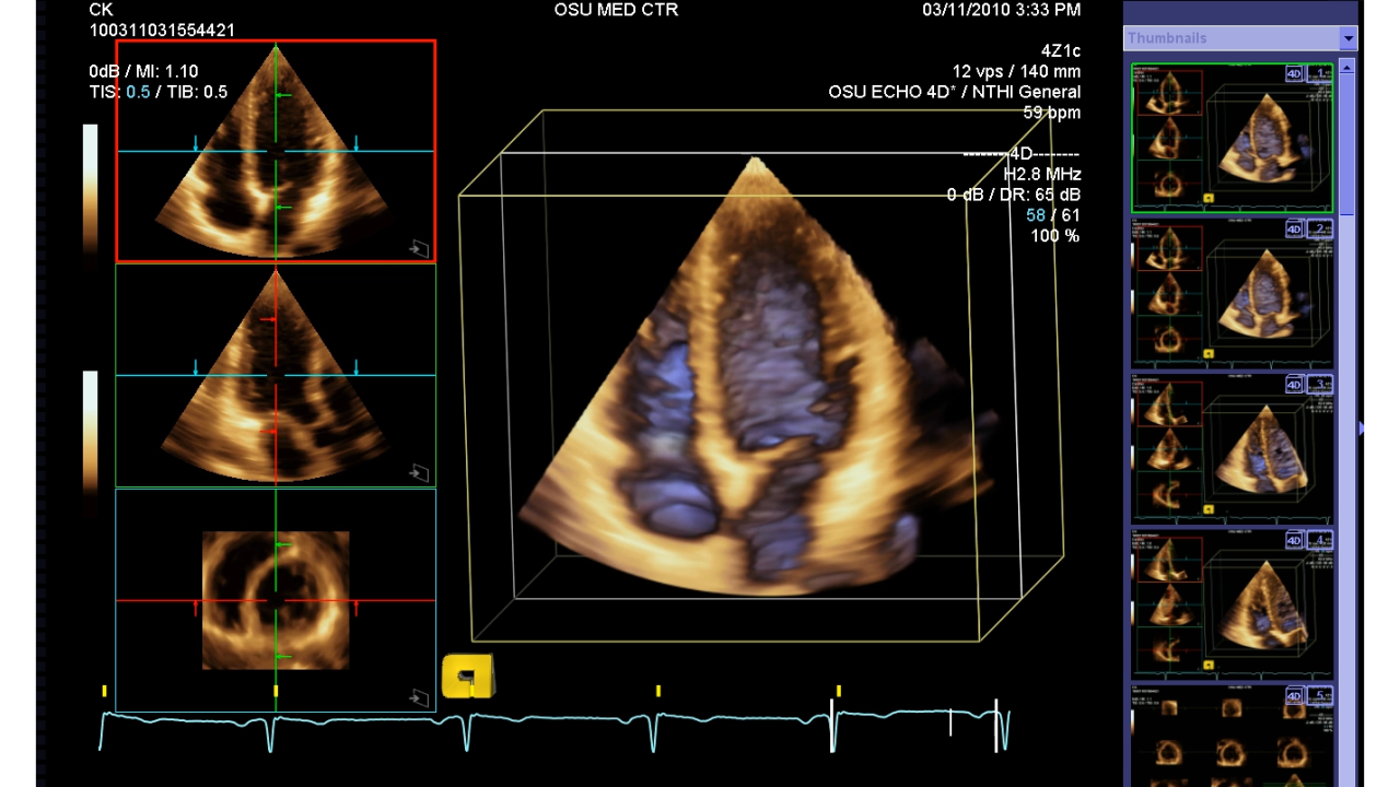 Model of 4D Left Ventricle4D model of left ventricle measuring ejection fraction, stroke volume and regional wall motion.
 Model of 4D Mitral Valve4D model of mitral valve made using Tomtec program.
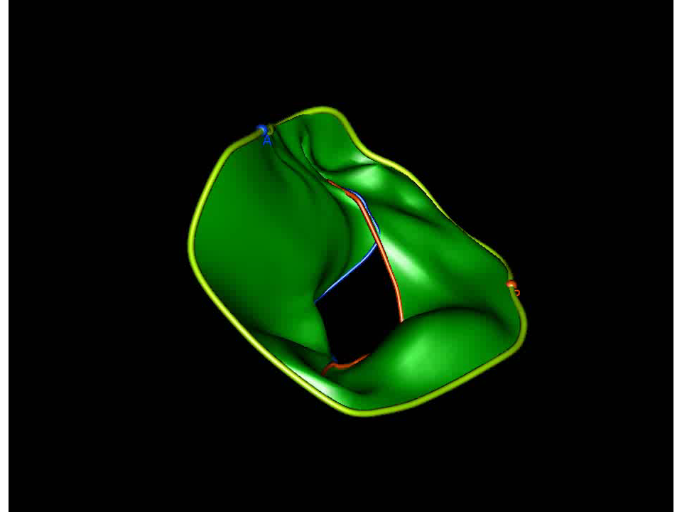 Model of 4D Mitral Valve with posterior prolapse4D model of mitral valve made using Tomtec program demonstrating posterior prolapse
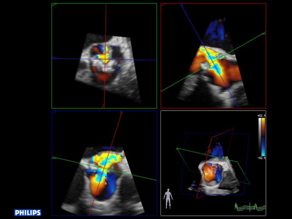 Multi-planar reconstruction of a 3D color datasetMulti-planar reconstruction of a 3D color dataset demonstrating measurement of the effective regurgitant oriface.
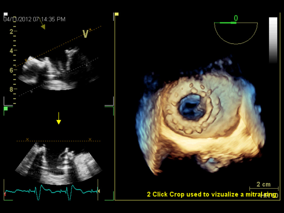 Post Mitral valve surgery on Real time 3D-TEEReal time 3D-TEE showing the mitral valve, post surgery from the left atrial perspective. The left panels show the…