The aortic valve is located between the left ventricle and the large vessel to the body which is called the aorta. Aortic valve stenosis is a condition in which the aortic valve leaflets are thickened and fused, making it harder for blood to flow from the ventricle to the aorta. Pressure rises in the left ventricle in order to pump blood through the narrowed valve. When the pressure build-up is significant, the muscle of the left ventricle becomes thickened, known as hypertrophy, and this can eventually lead to muscle damage.
In infants and children a catheter procedure can be performed to open the valve and improve blood flow. This procedure is called a transcatheter balloon valvuloplasty. A balloon catheter is threaded from the large artery in the groin into the aorta and through the narrowed aortic valve. The balloon on the end of the catheter is then inflated to stretch, or tear, the aortic valve leaflets.
Once the catheter is removed, blood can flow more easily through the valve and the stenosis is relieved. Because the balloon valvuloplasty often tears the aortic valve leaflets they may not close tightly, causing leakage of blood backward into the ventricle, also known as insufficiency or regurgitation.
Over time, the valve can become narrowed again or the regurgitation can progress, thus many patients who have had a transcatheter balloon valvuloplasty require surgery or another catheter intervention later in life.
©2026 Medmovie.com. All rights reserved. Medmovie.com creates and licenses medical illustrations and animations for educational use. Our goal is to increase your understanding of medical terminology and help communication between patients, caregiver and healthcare professionals. The content in the Media Library is for your information and education purposes only. The Media Library is not a substitute for professional medical advice, diagnosis or treatment for specific medical conditions.
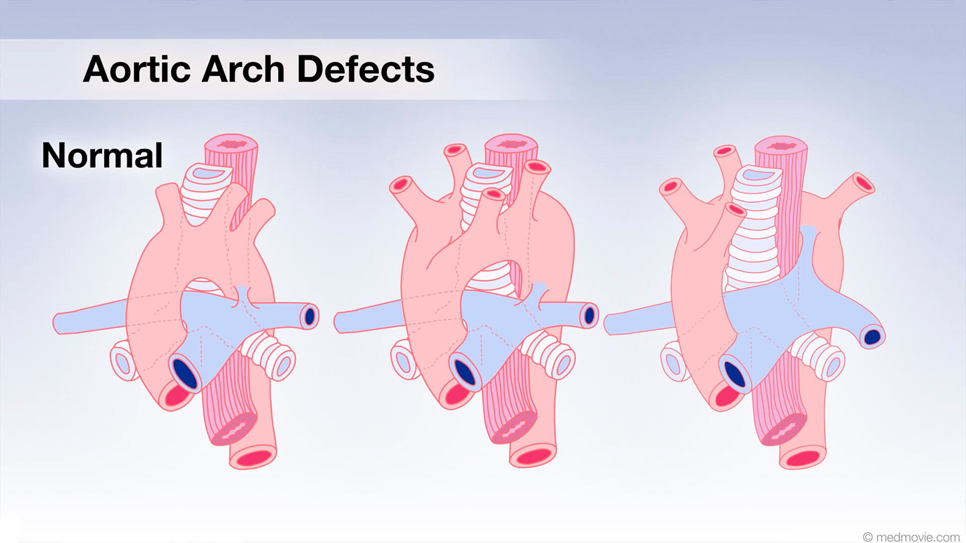 Aortic Arch DefectsIn the early stages of fetal development, two aortic arches come from the heart, ascend upward and then descend behind…
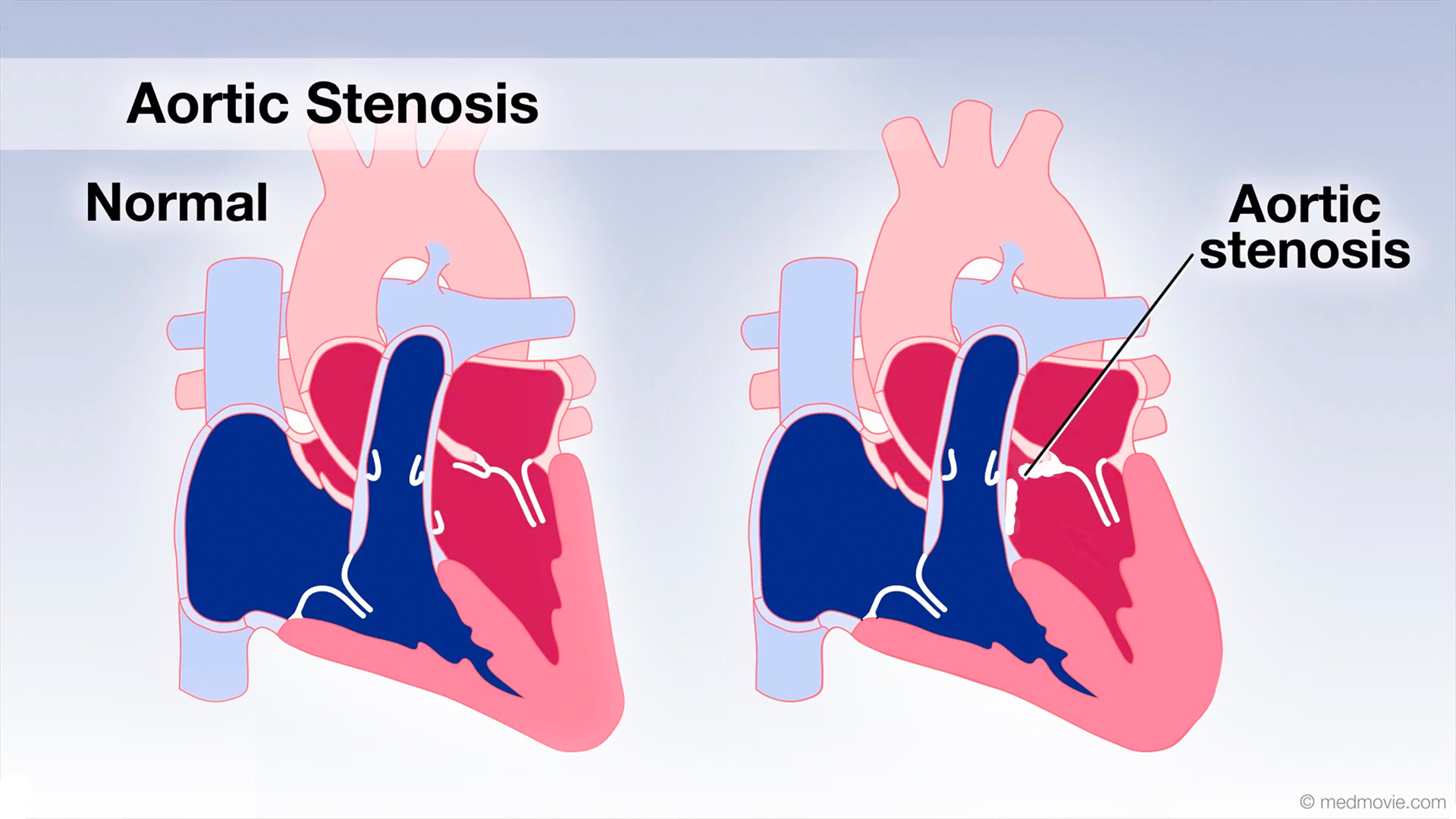 Aortic Valve StenosisThe aortic valve is located between the left ventricle and the large vessel to the body which is called the aorta.…
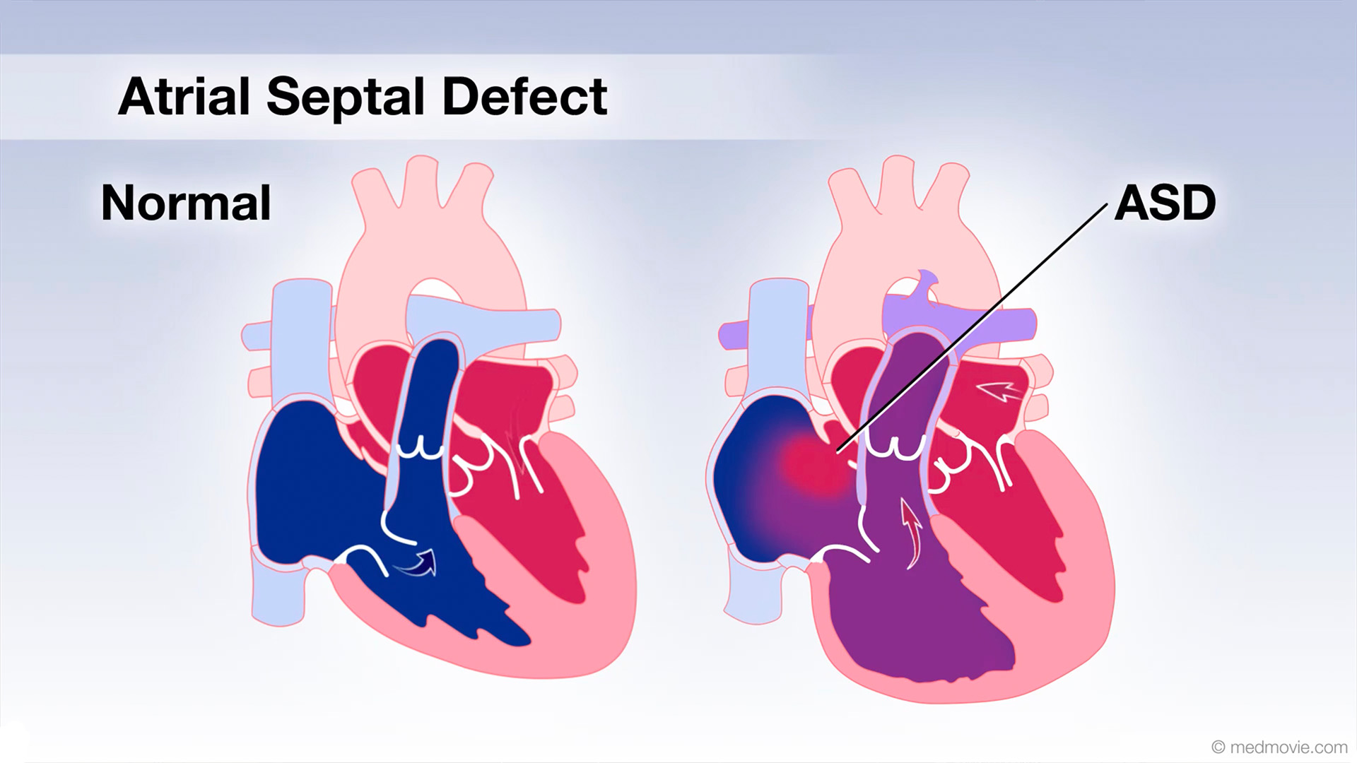 Atrial Septal DefectAn atrial septal defect, or ASD, is a hole in the wall that separates the upper two chambers of the heart, known as the…
 AV CanalThe atrioventricular canal, or AV canal, is the structure that forms the center of the heart. During normal fetal…
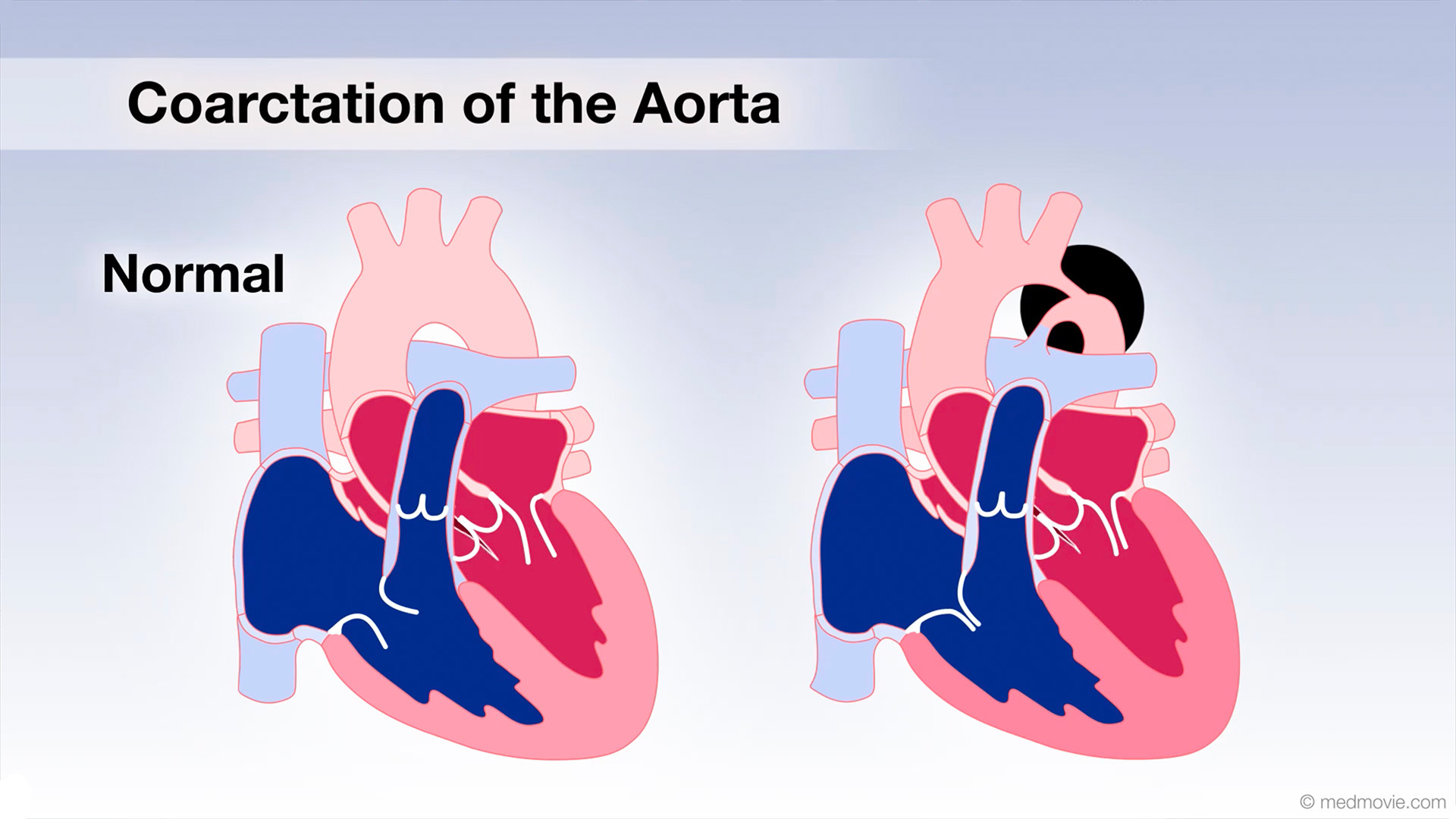 Coarctation of AortaCoarctation of the Aorta is a congenital defect in which the upper part of the descending aorta is severely narrowed or…
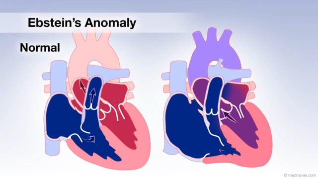 Ebstein's AnomalyThe tricuspid valve is the valve between the right atrium and the right ventricle, and in Ebstein’s anomaly it is…
 Heartbeat - ElectricHeart muscle cells generate electrical signals that are conducted through the heart by special fibers which stimulate…
 Heart Lung BypassA heart-lung -or cardiopulmonary bypass - machine is used during heart surgery to deliver oxygenated blood to the body…
 Hypoplastic Left Heart SyndromeHypoplastic left heart syndrome is the result of underdevelopment of the left heart structures and includes a tiny, or…
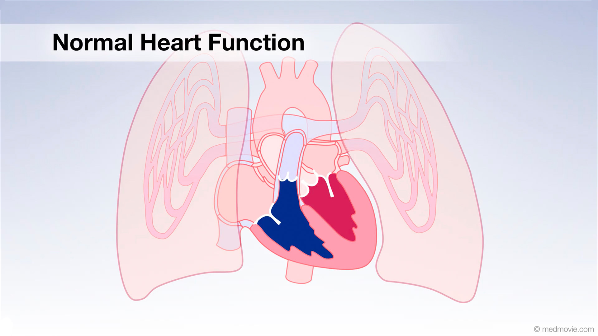 Normal Heart FunctionBlood flows through the heart in a specific pattern in order to pump deoxygenated, or blue blood to the lungs and…
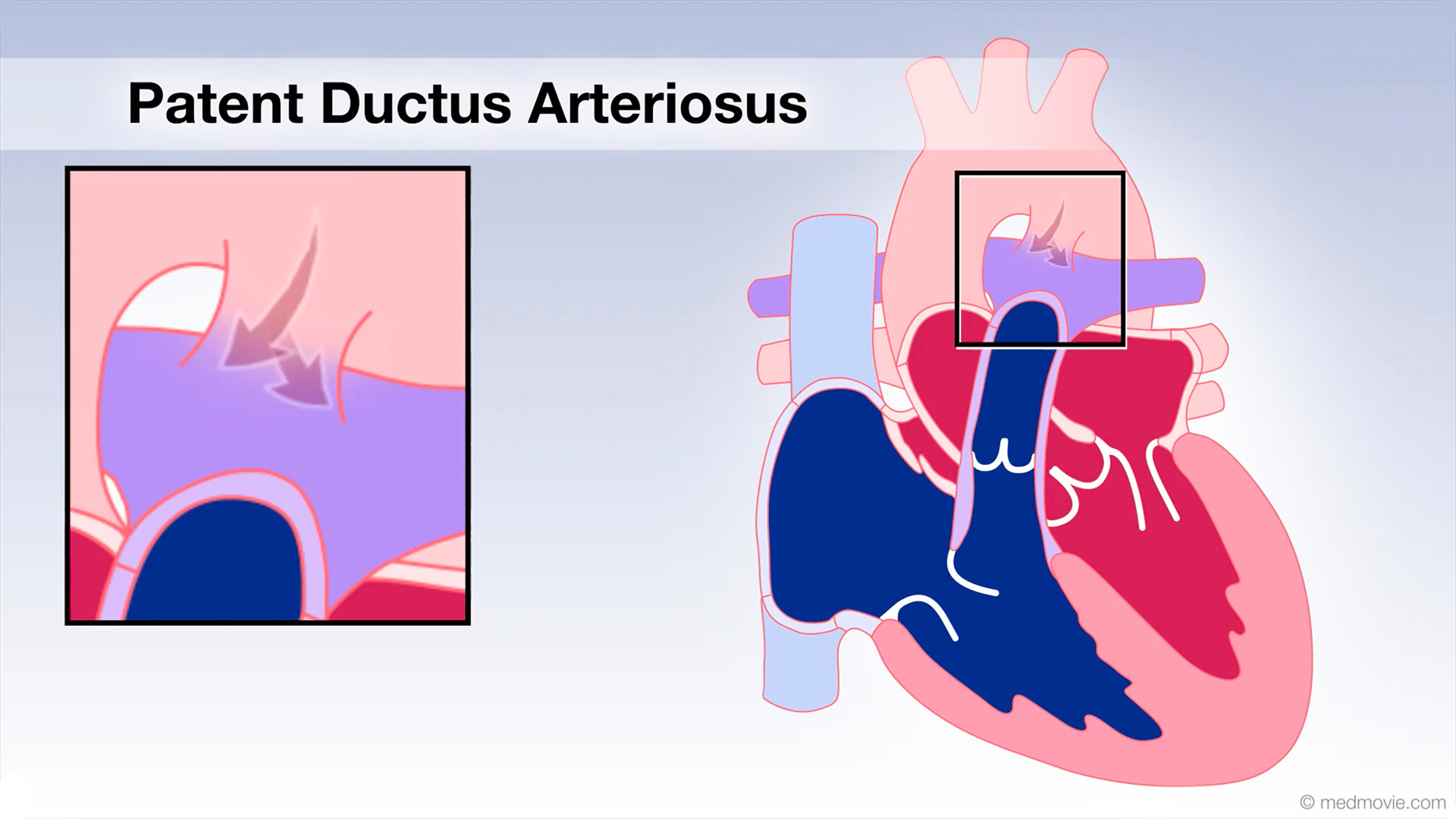 Patent Ductus Arter.The ductus arteriosus is a blood vessel that connects the blood vessel going to the lungs, known as the pulmonary…
 Pulm. Valve StenosisThe pulmonary valve is located between the right ventricle and the large blood vessel to the lungs which is called the…