
Stock Illustration and Animation Samples
We have a large collection of stock illustration available for licensing. These are just a few samples of our stock collection. Please use our search feature. If you cannot find what you are looking for, please contact us.
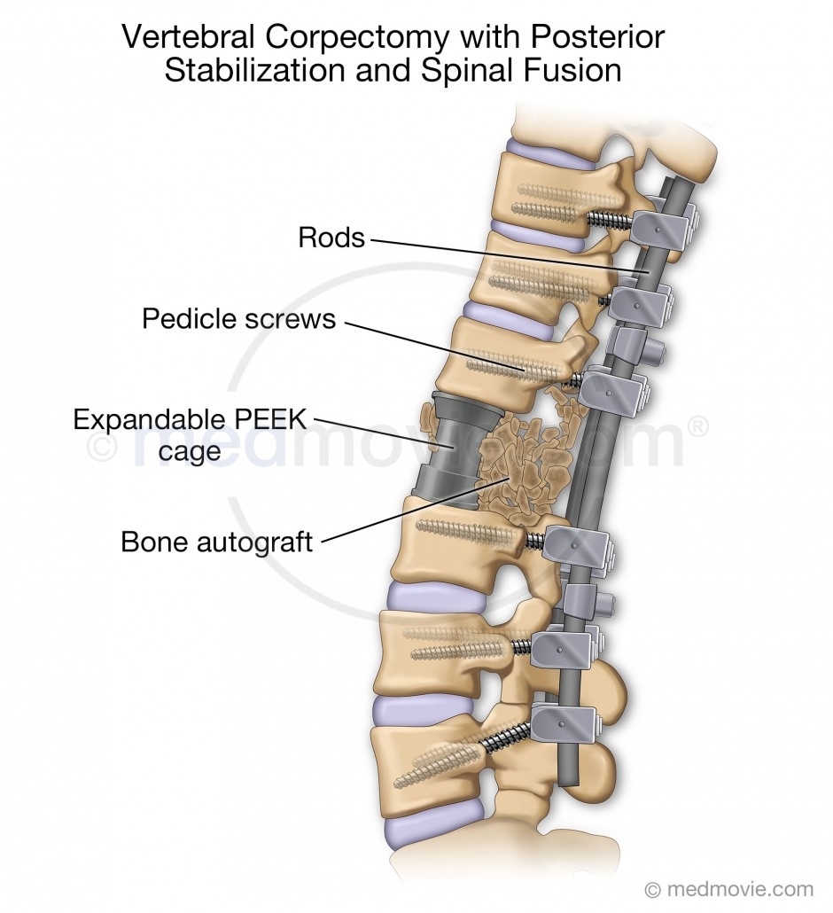
This illustration depicts posterior spine stabilization and spinal fusion with a vertebral corpectomy, as seen from the side.
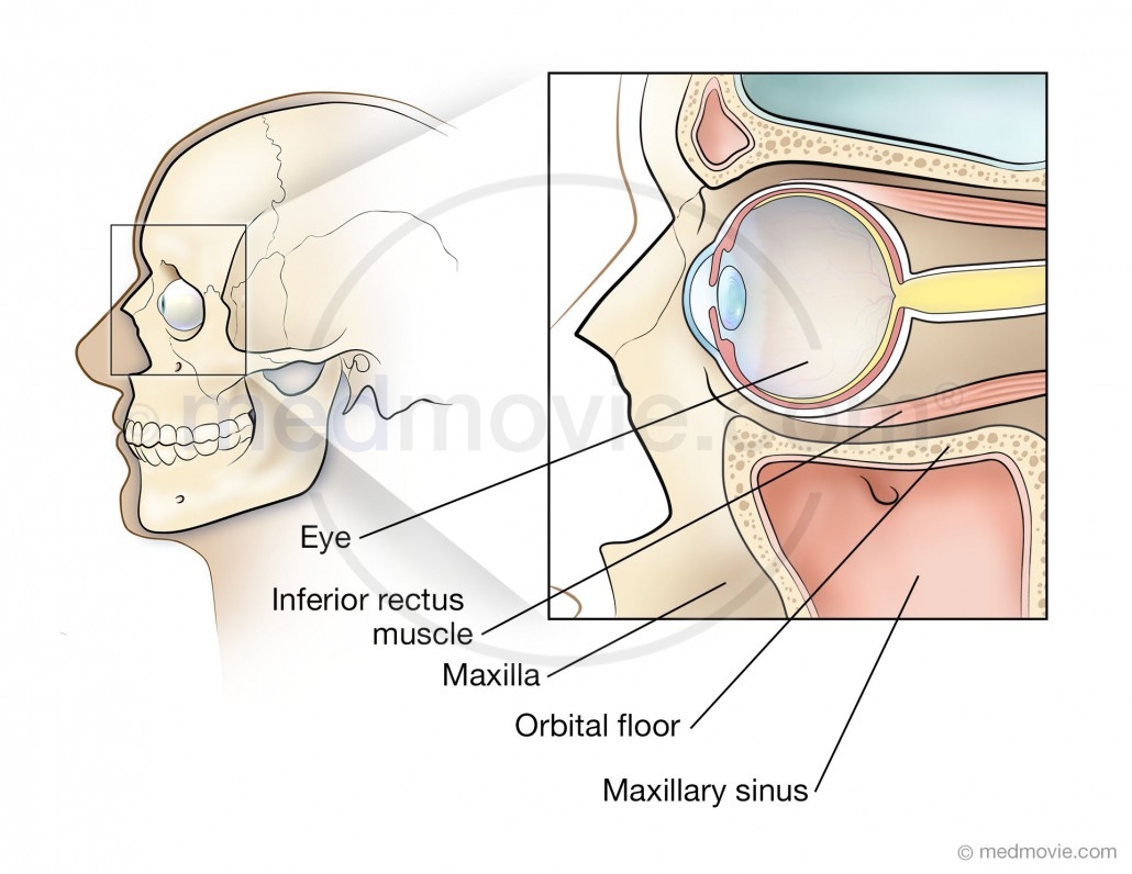
Illustration depicting a cross-section of the orbit of the eye.
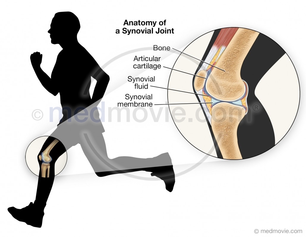
This illustration depicts the knee joint both in the context of a silhouetted figure running, and as a close-up illustration. Labels highlight synovial joint structures.
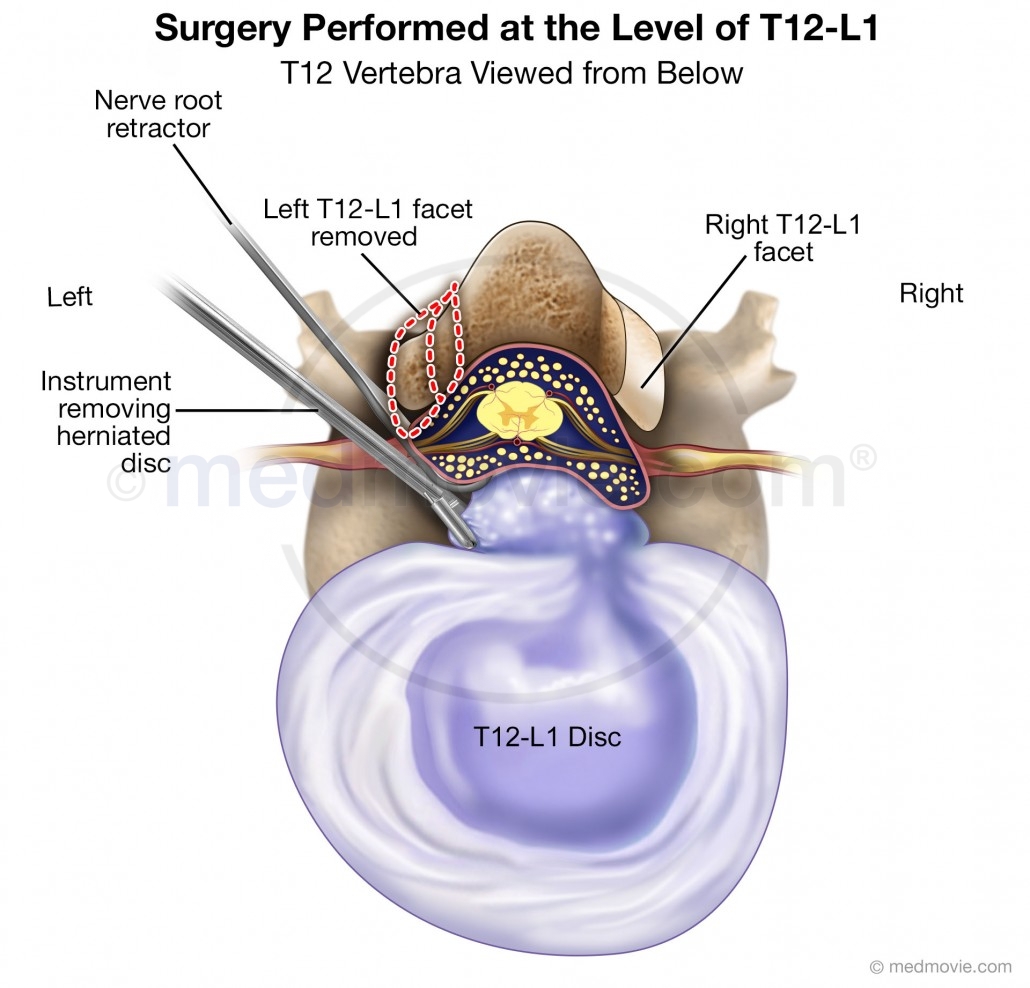
This illustration depicts a herniated disc surgery, repairing the T12-L1 disc, as viewed from below.
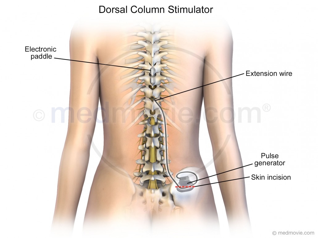
This illustration depicts the placement of a dorsal column stimulator in the body of an adult female, as seen from the posterior / back side.
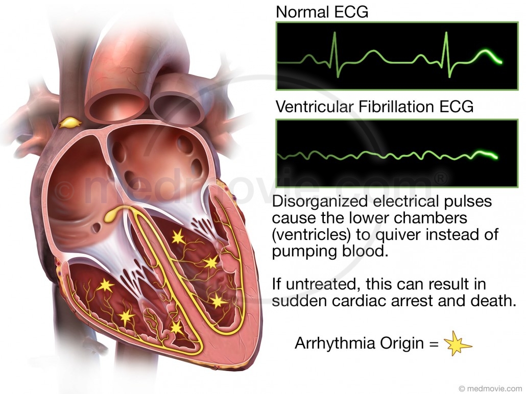
Illustration of a heart with ventricular fibrillation including a normal ECG compared with an abnormal ECG.

This illustration depicts, with a female figure, the best way to get up and sit down on a chair after receiving an artificial joint; it is best to keep the artificial leg out in front.
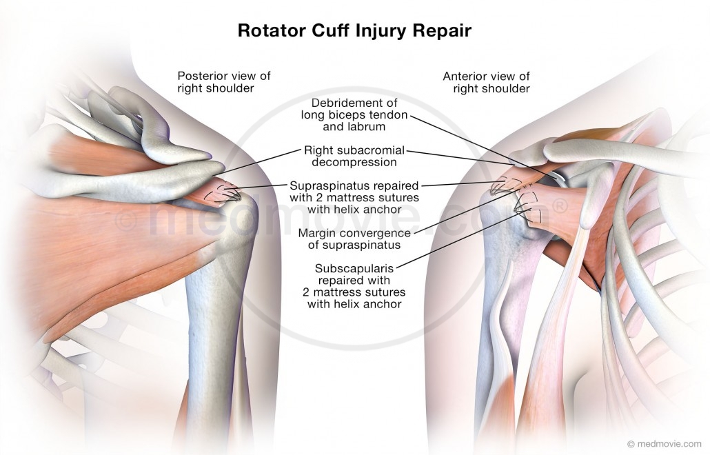
This illustration depicts the finished surgical repairs to a rotator cuff injury, seen from both anterior and posterior views and including labels.
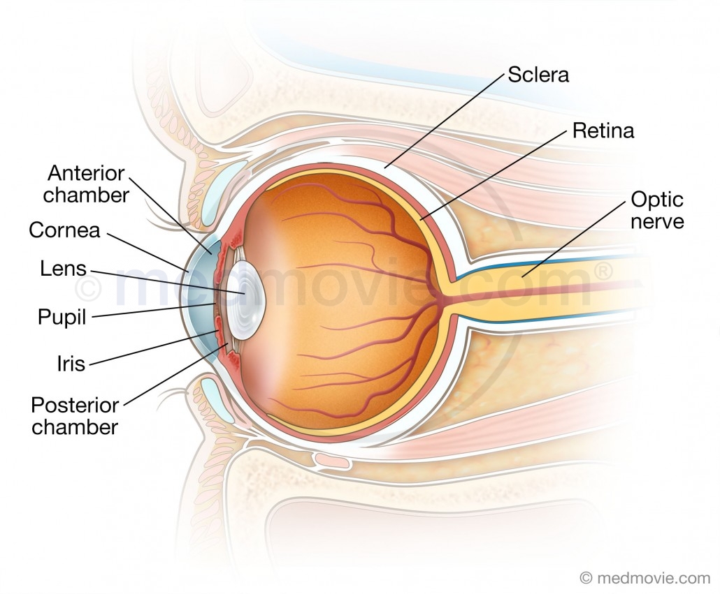
Anatomy of the Eye
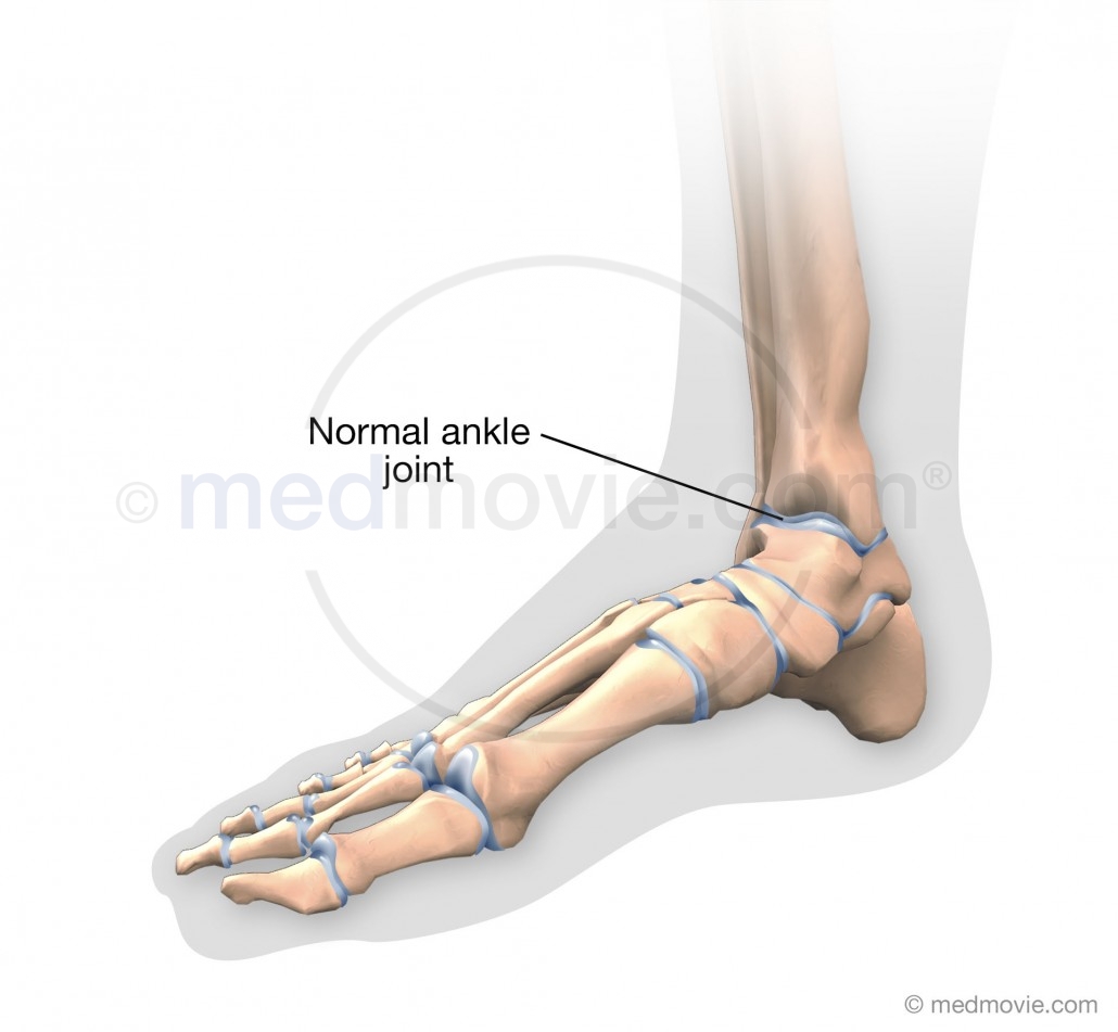
This illustration depicts a normal ankle joint.

This Illustration depicts an elbow joint replacement.

This illustration depicts the internal abdominal organs of a baby including the esophagus, stomach, liver, spleen, small intestine, and large intestine.
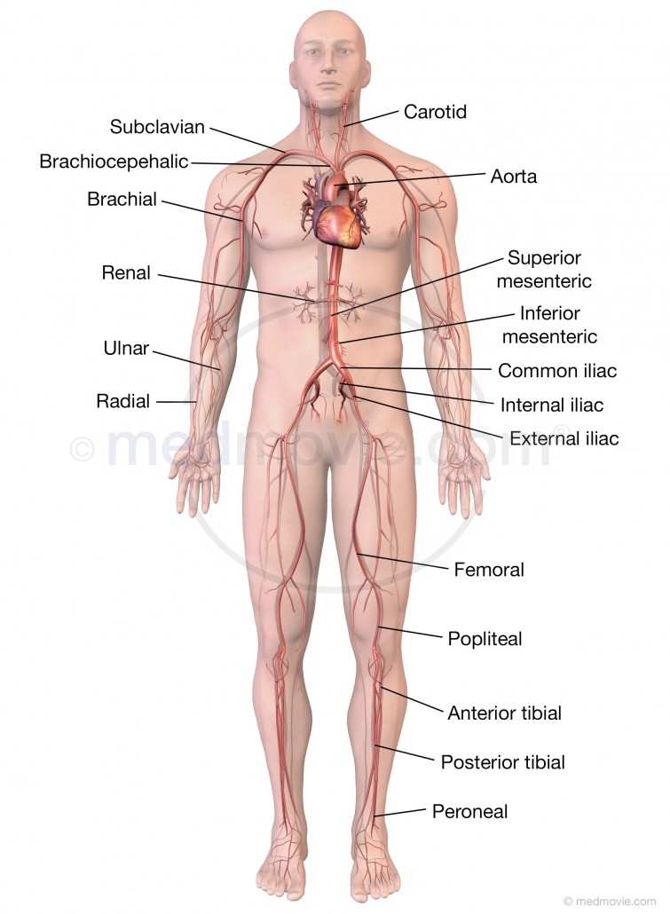
Illustration of the major arteries of the body.
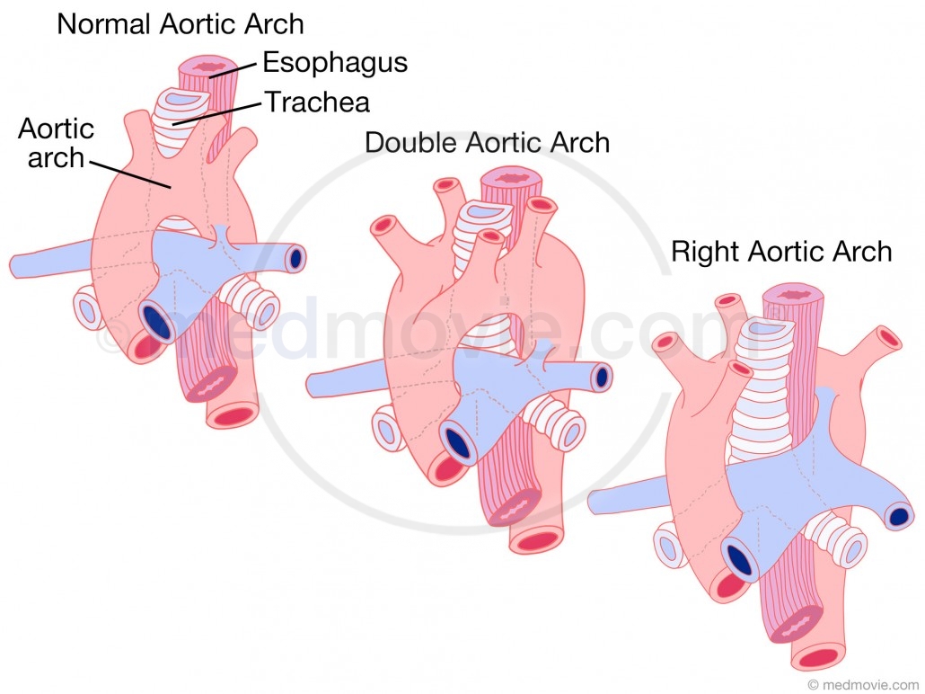
Illustration depicting a normal aortic arch, a double aortic arch, and a right aortic arch.
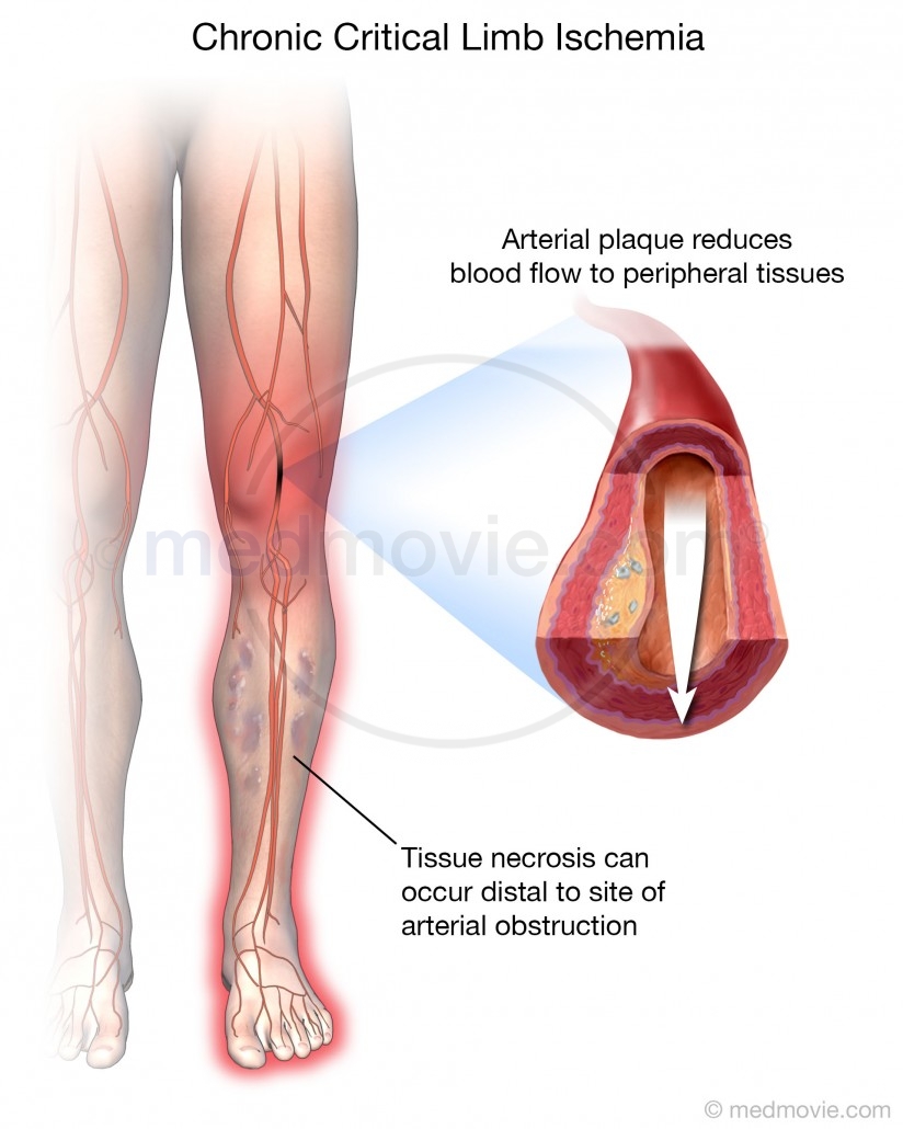
This illustration depicts an arterial plaque reducing blood flow in a peripheral artery. This causes ischemia in the distal tissue and can cause tissue necrosis.
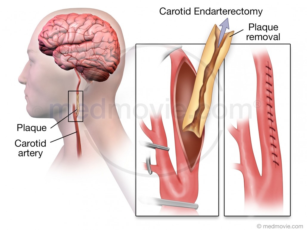
Illustration of endarterectomy surgery being performed on the carotid artery with plaque removal.
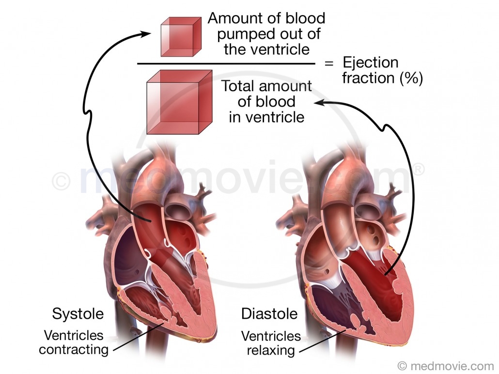
This illustration depicts the ejection fraction of the heart and how it is calculated from the amount of blood that is pumped out of the ventricle divided by the total amount of blood in the ventricle.
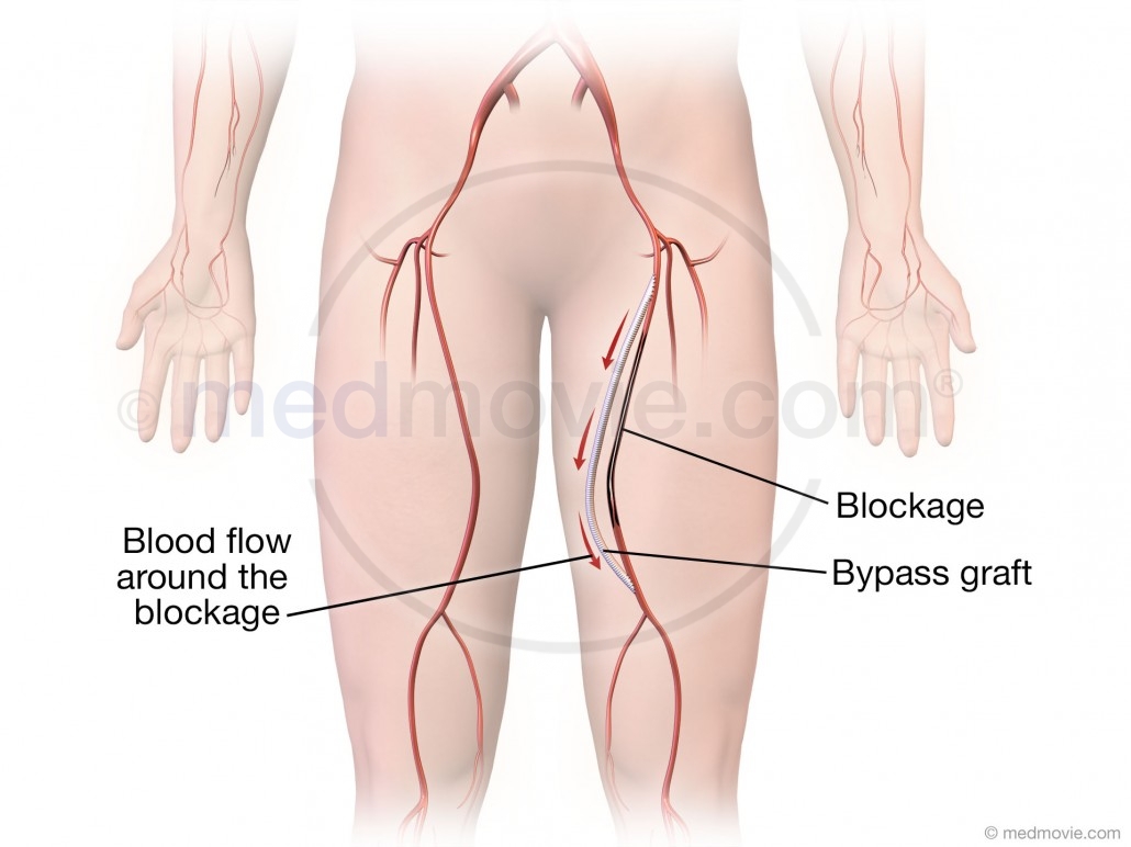
Illustration depicting the placement of a bypass graft on the femoral artery of the leg.
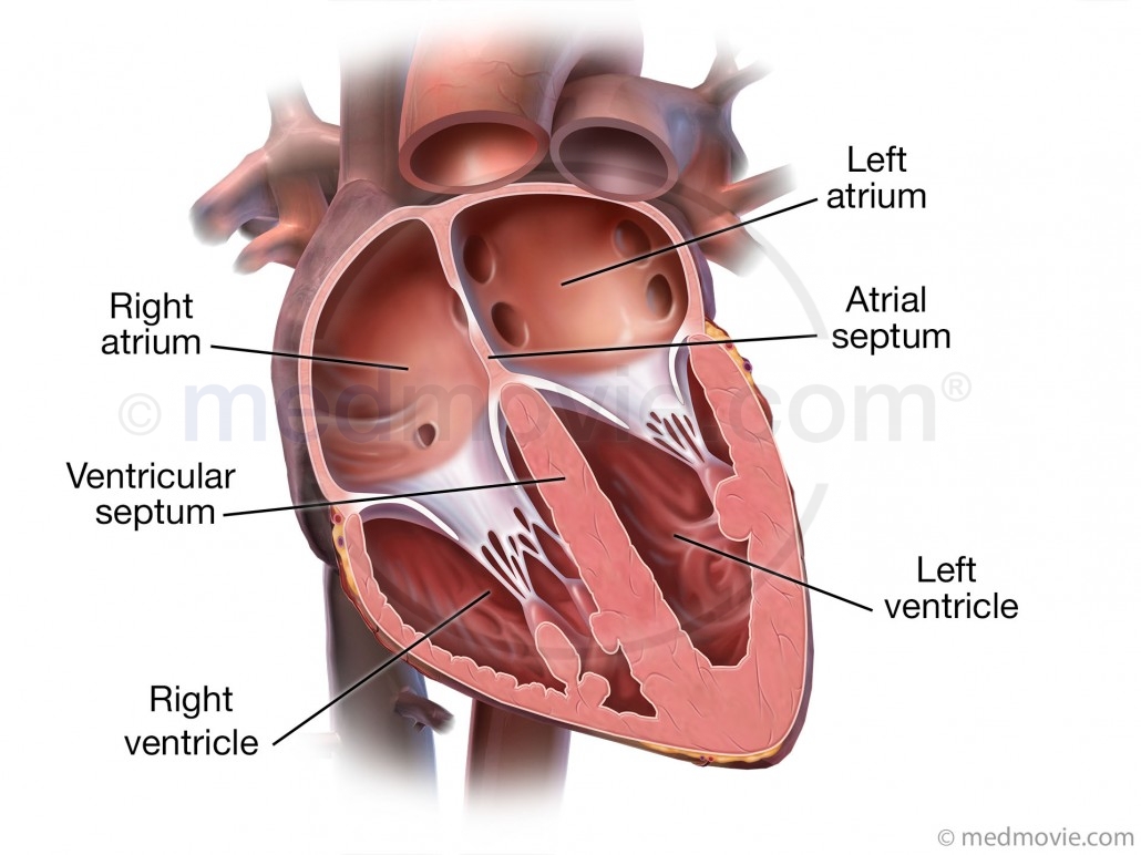
Illustration of a cross section of the heart with labels for the atria and ventricles of the heart.

Illustration of a heart with a single ventricle compared to a normal heart.