Hypoplastic left heart syndrome is the result of underdevelopment of the left heart structures and includes a tiny, or hypoplastic, left ventricle, complete closure or severe narrowing of the aortic and mitral valves, an abnormally small aorta, a patent foramen ovale and a large patent ductus arteriosus, or PDA. The severe underdevelopment of the left heart structures prevents oxygen-rich blood from flowing to the body normally. Thus, the right heart takes over and pumps blood to the body through the PDA. A patent foramen ovale is a hole between the upper chambers of the heart, or the atria, that allows oxygen-rich blood from the lungs to bypass the left heart by flowing from the left atrium to the right atrium. In this way, oxygen-rich blood reaches the right heart, PDA, and the aorta to supply the body with oxygen.
This condition can be improved by a series of three operations. The first operation, which is called the Norwood procedure, is done shortly after birth. After placing the baby on the heart-lung machine, the aorta is opened and the PDA is tied closed. The pulmonary artery is divided into two parts. The part of the pulmonary artery that is close to the heart is sewn to the small aorta with a patch to make it bigger. This allows blood from the right ventricle to flow easily into the aorta to pump blood to the body. In order to allow blood to flow to the lungs a small tube, known as a modified Blalock-Taussig shunt, is placed between a branch of the aorta and the pulmonary arteries. Sometimes, instead of a Blalock-Taussig shunt, a Sano conduit is placed between the right ventricle and the pulmonary arteries.
At the second operation, known as the bidirectional Glenn shunt or the hemi-Fontan, the shunt or conduit is closed off and the large vein draining the head and arm, known as the superior vena cava, is connected directly to the pulmonary arteries. At the third operation, or Fontan, the large vein draining the lower part of the body, known as the inferior vena cava, is connected to the pulmonary arteries. At the end of the three operations, the oxygen-poor blood from the veins flows directly into the lungs and the oxygen-rich blood from the lungs flows into the left atrium to the right ventricle and is pumped to the body.
©2026 Medmovie.com. All rights reserved. Medmovie.com creates and licenses medical illustrations and animations for educational use. Our goal is to increase your understanding of medical terminology and help communication between patients, caregiver and healthcare professionals. The content in the Media Library is for your information and education purposes only. The Media Library is not a substitute for professional medical advice, diagnosis or treatment for specific medical conditions.
 Aortic Arch DefectsIn the early stages of fetal development, two aortic arches come from the heart, ascend upward and then descend behind…
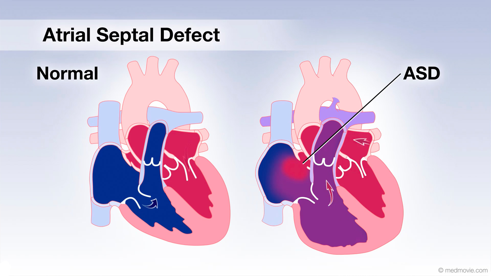 Atrial Septal DefectAn atrial septal defect, or ASD, is a hole in the wall that separates the upper two chambers of the heart, known as the…
 AV CanalThe atrioventricular canal, or AV canal, is the structure that forms the center of the heart. During normal fetal…
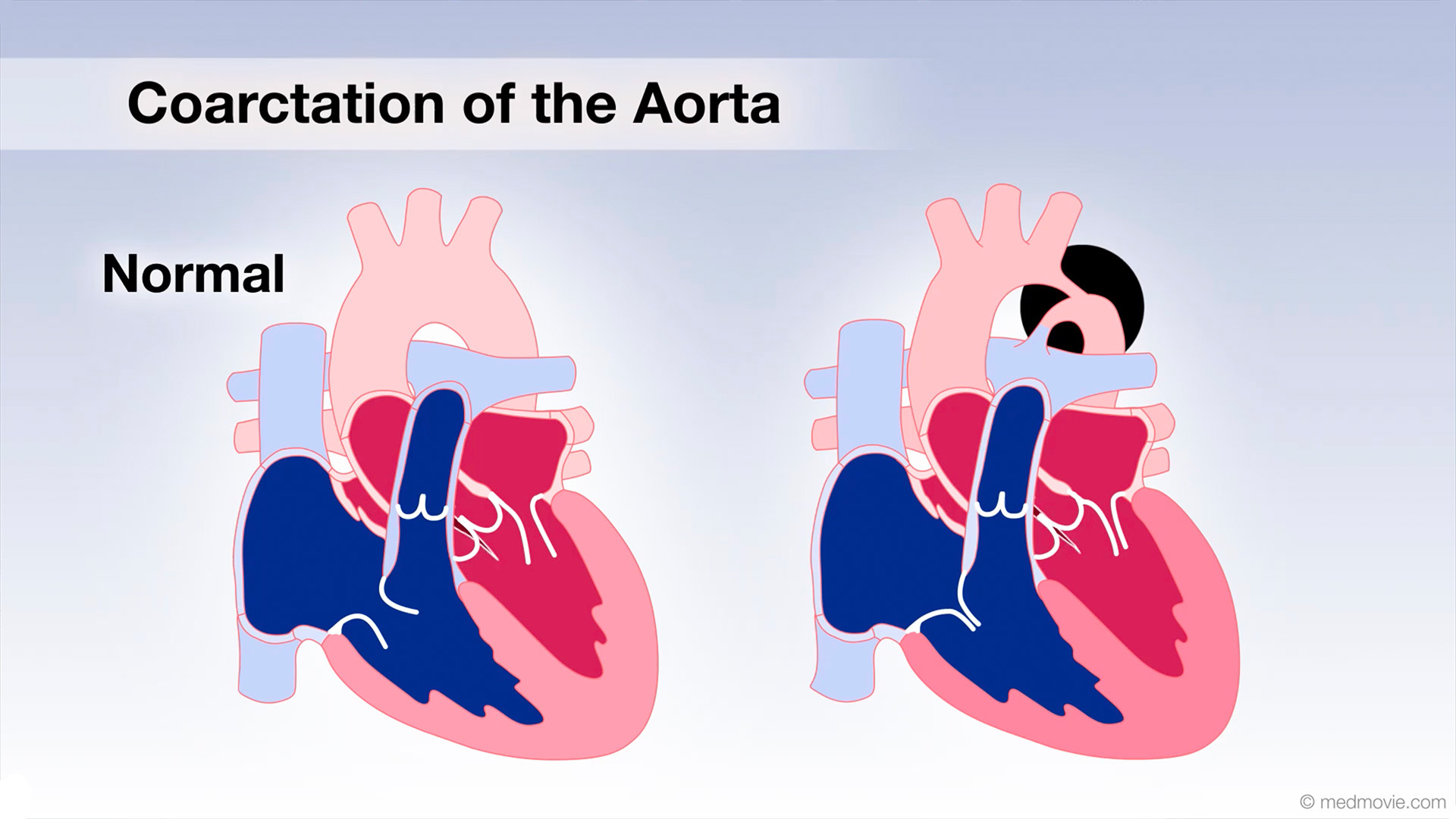 Coarctation of AortaCoarctation of the Aorta is a congenital defect in which the upper part of the descending aorta is severely narrowed or…
 Hypertrophic CardiomyopathyThe normal heart has strong muscular walls that pump blood out of the heart.
Hypertrophic cardiomyopathy is a…
Hypertrophic CardiomyopathyThe normal heart has strong muscular walls that pump blood out of the heart.
Hypertrophic cardiomyopathy is a…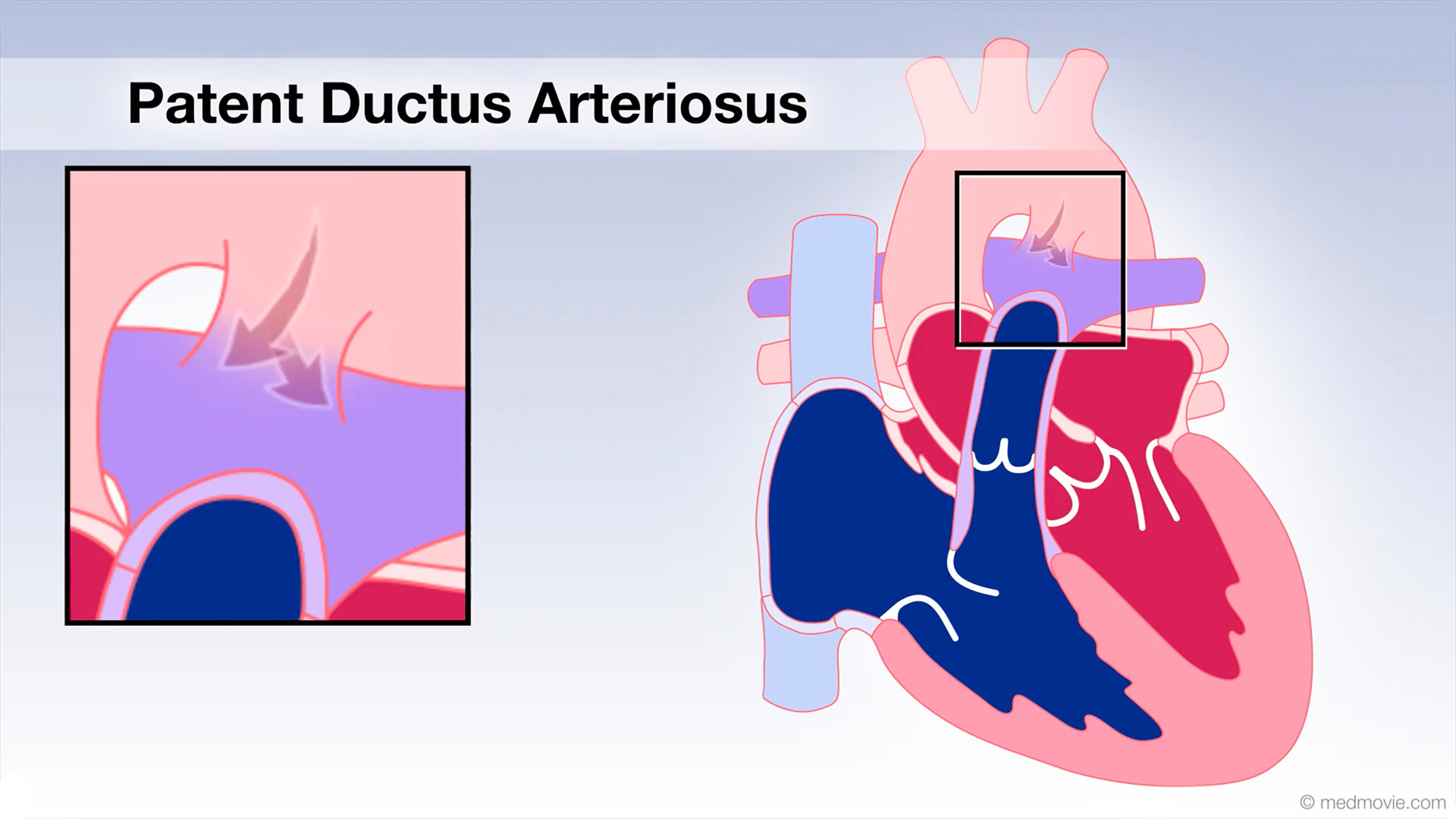 Patent Ductus Arter.The ductus arteriosus is a blood vessel that connects the blood vessel going to the lungs, known as the pulmonary…
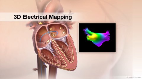 3D Electrical MappingElectrical mapping of the heart is a procedure that is used to diagnose the origins of arrhythmias. This procedure uses…
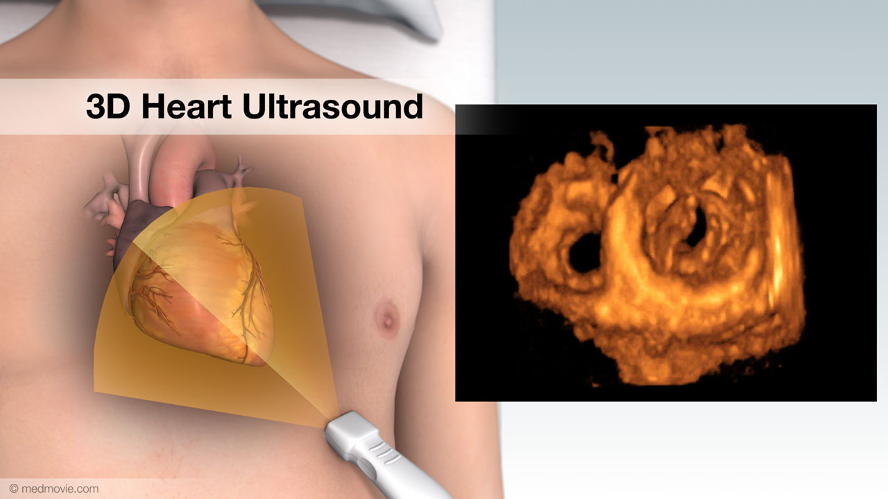 3D Heart UltrasoundA 3D heart ultrasound, also known as a 3D echocardiogram, is a diagnostic test that creates 3D videos of the heart.…
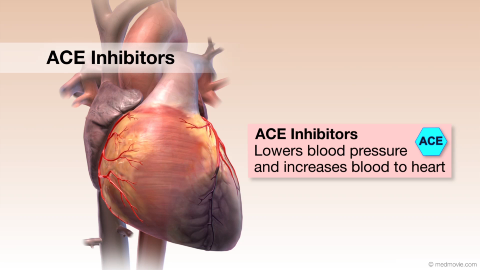 ACE InhibitorsACE Inhibitors (Angiotensin-Converting Enzyme) Inhibitors are drugs used to treat high blood pressure and heart failure…Aortic Arch DefectsIn the early stages of fetal development, two aortic arches come from the heart, ascend upward and then descend behind…
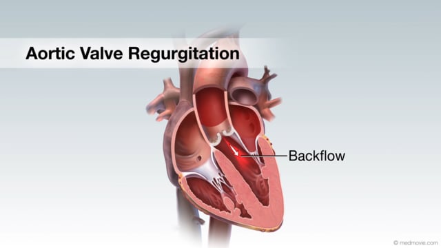 Aortic Valve RegurgitationAortic valve regurgitation results when the aortic valve, which sits between the left ventricle and the aorta, cannot…
 Aortic Valve StenosisThe aortic valve is located between your heart’s main pumping chamber (left ventricle) and the major artery (aorta)…
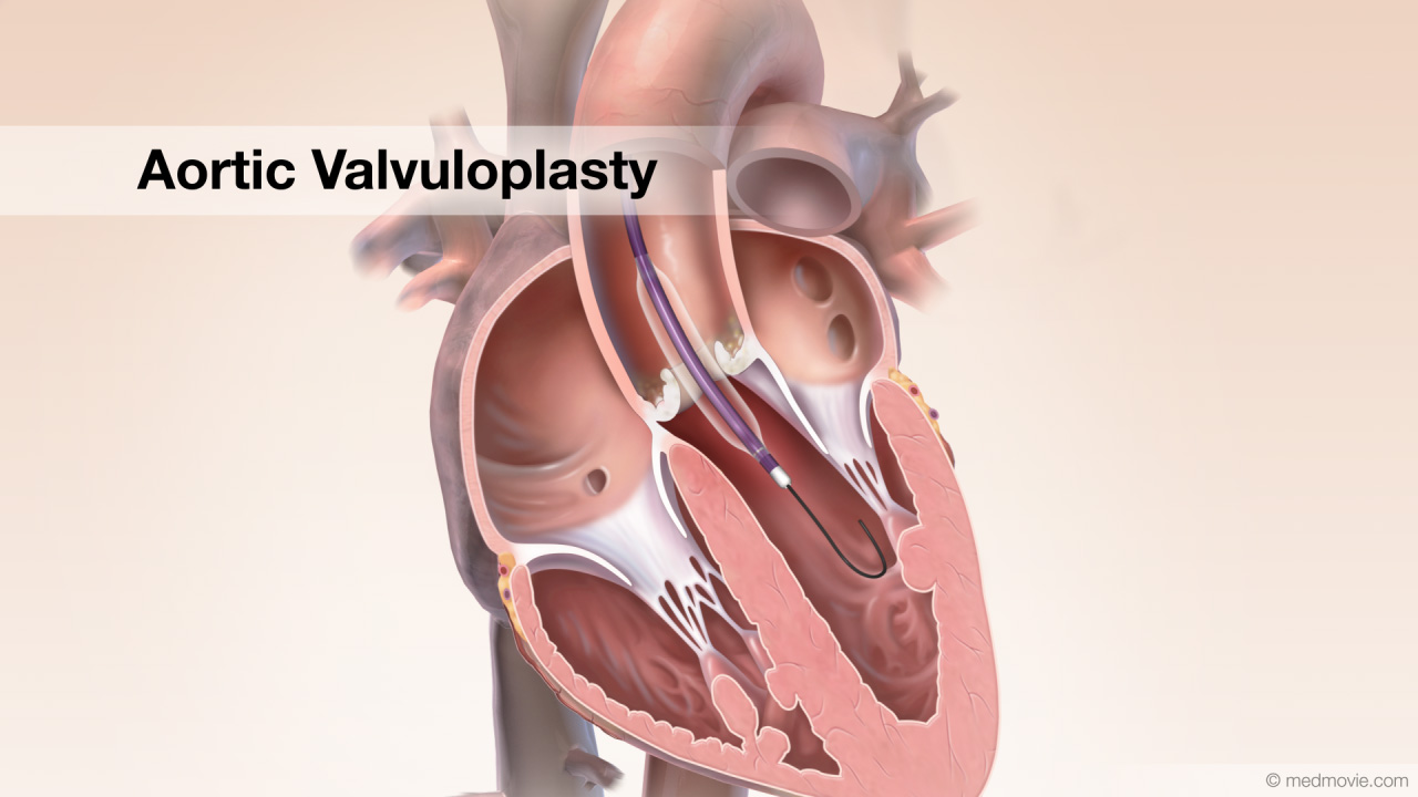 Aortic ValvuloplastyStenotic aortic valve disease can require a procedure called aortic valvuloplasty. Calcific aortic stenosis is shown…
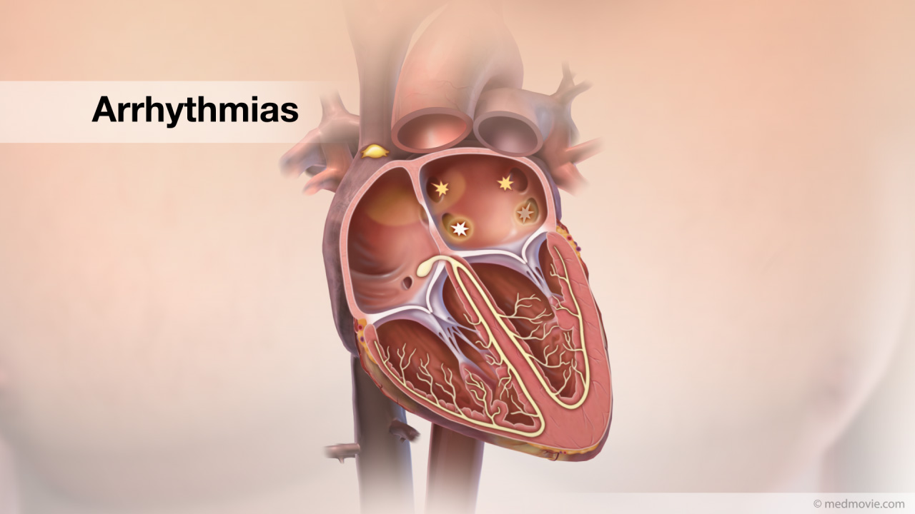 ArrhythmiasThe heartbeat is controlled by the electrical system of the heart. This system is made up of several sequences that…
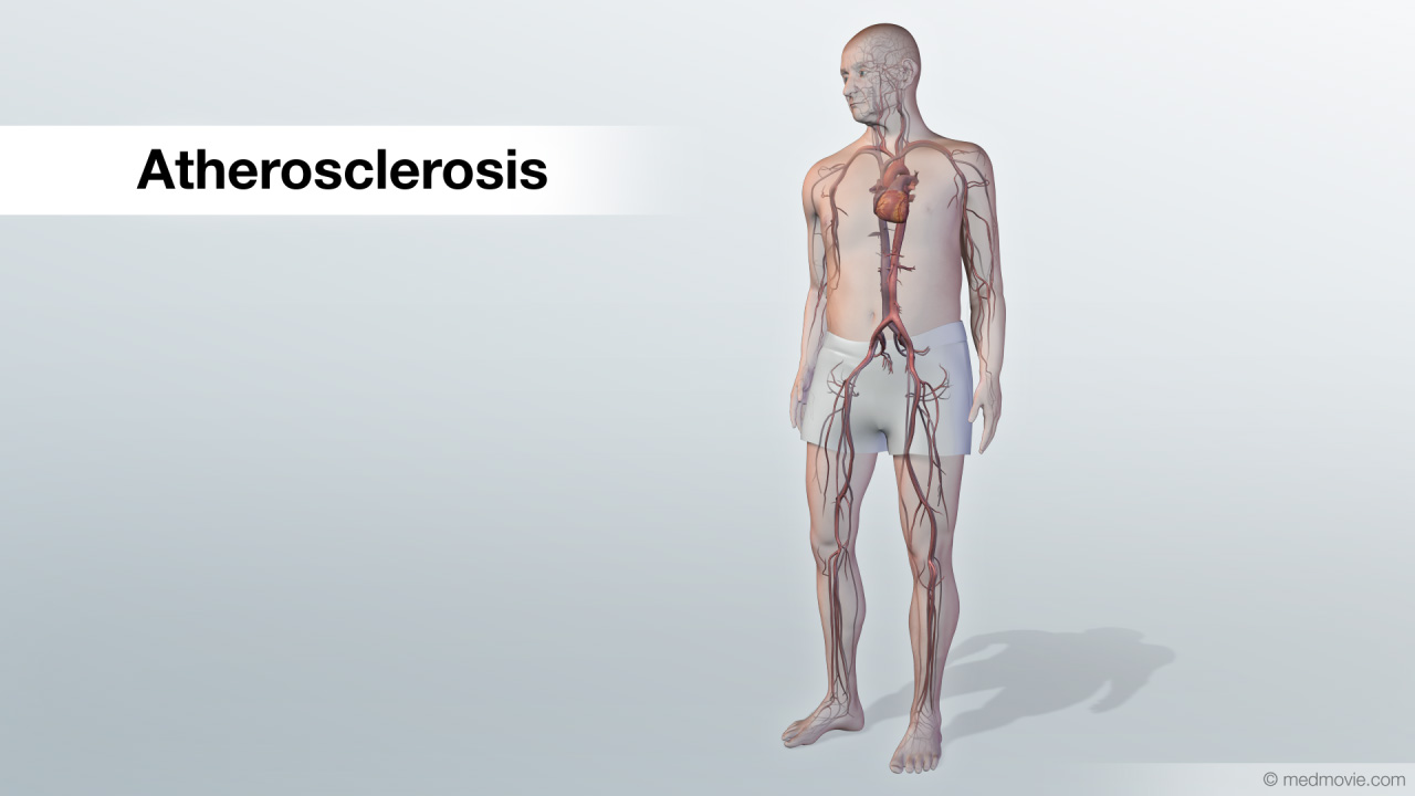 AtherosclerosisAtherosclerosis is a disease that affects the arteries, which carry blood to the organs of the body.
Normal arteries…
AtherosclerosisAtherosclerosis is a disease that affects the arteries, which carry blood to the organs of the body.
Normal arteries…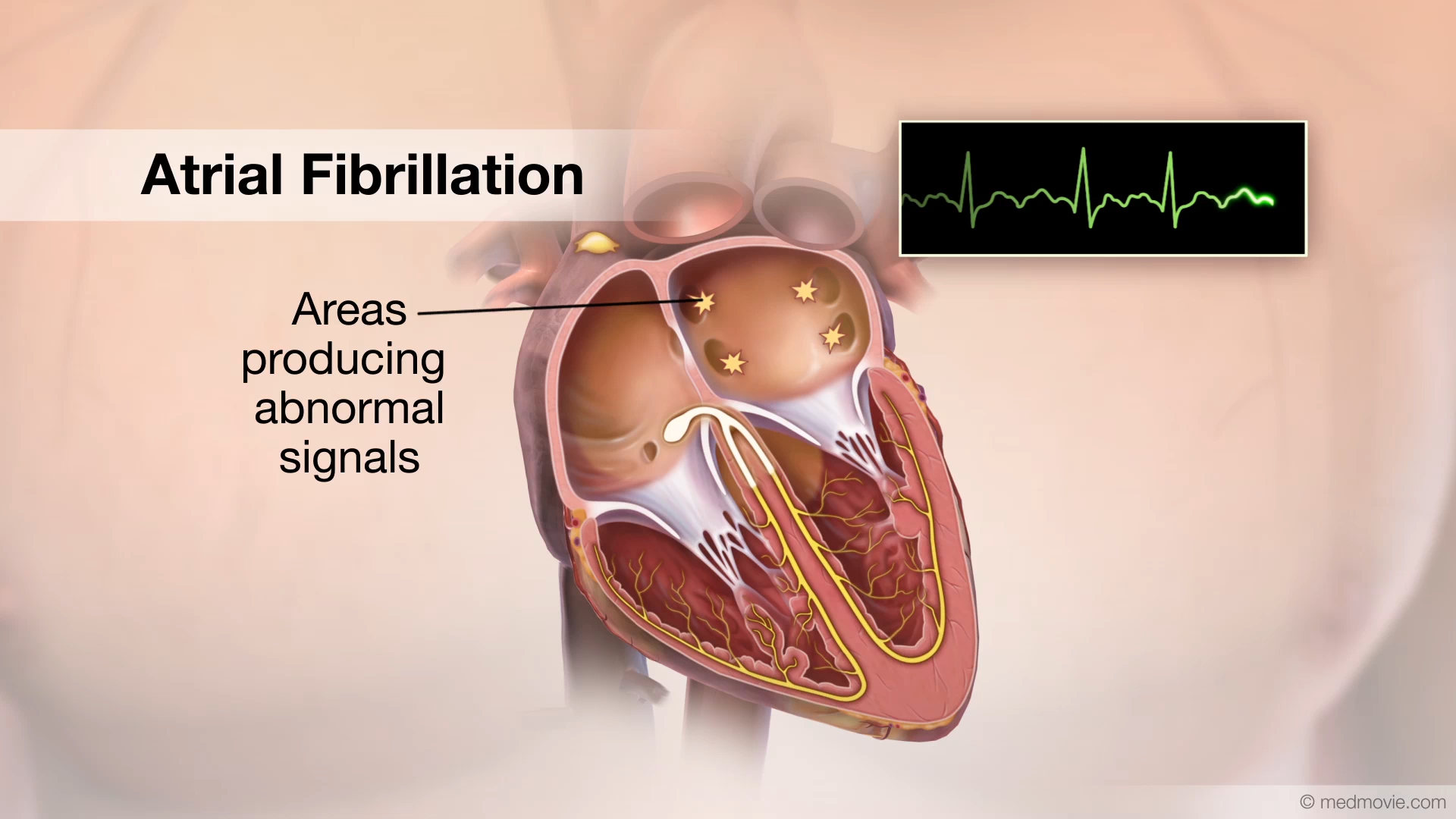 Atrial FibrillationThe heartbeat is controlled by the electrical system of the heart. This system is made up of several parts that tell…
 Atrial Fibrillation AblationAtrial fibrillation is a heart arrhythmia in which abnormal electrical signals begin in the atria (top chambers) Atrial…
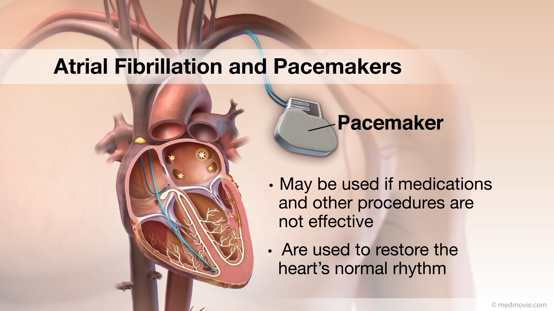 Atrial Fibrillation PacemakersAtrial fibrillation is a heart arrhythmia in which abnormal electrical signals begin in the atria (top chambers) of the…
 Atrial Fibrillation CardioversionAtrial fibrillation is the most common heart rhythm disorder. In atrial fibrillation, abnormal signals from the left…
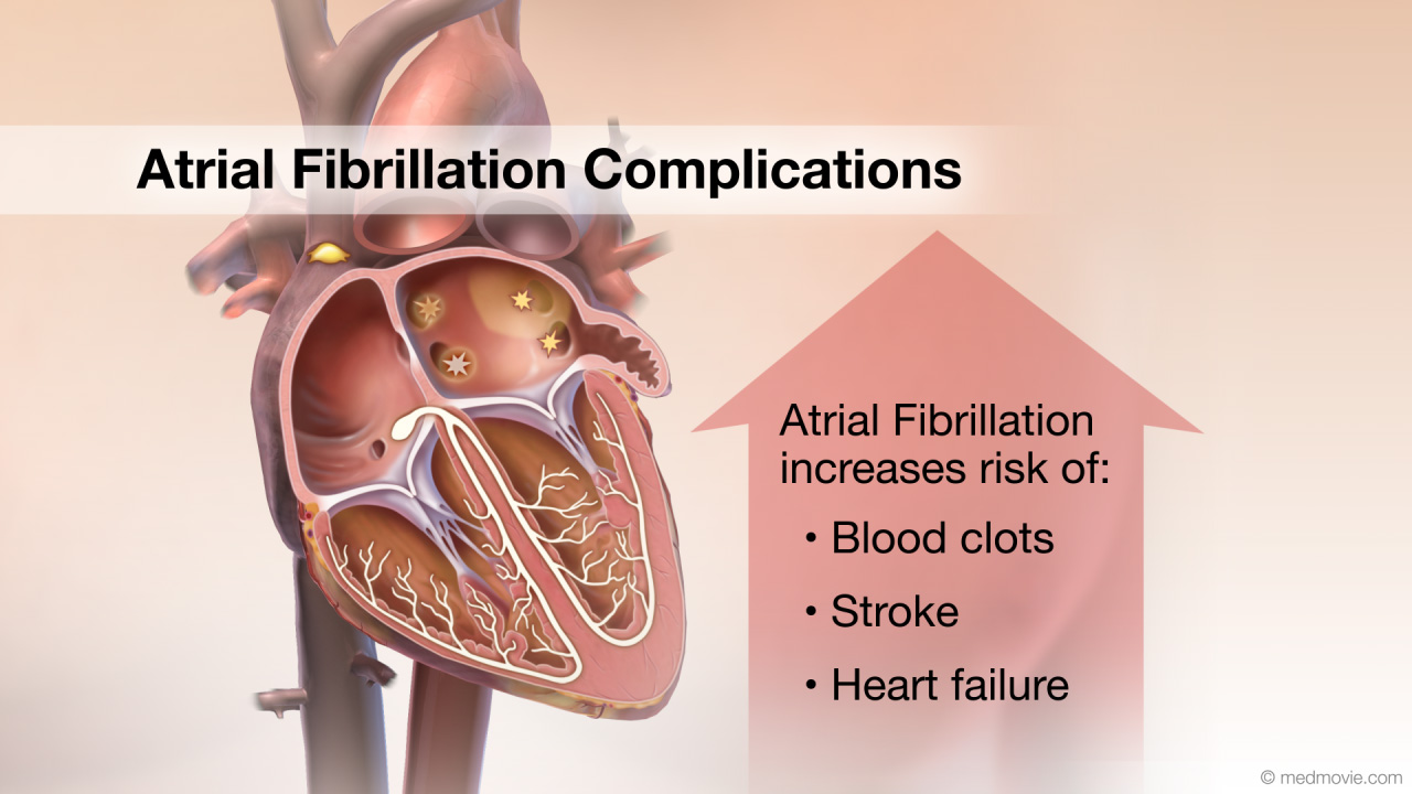 Atrial Fibrillation ComplicationsAtrial fibrillation (AF) is a heart arrhythmia in which abnormal electrical signals begin in the atria (top chambers)…
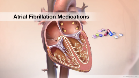 Atrial Fibrillation MedicationsAtrial Fibrillation is a heart arrhythmia in which abnormal electrical signals begin in the atria (or top chambers) of…
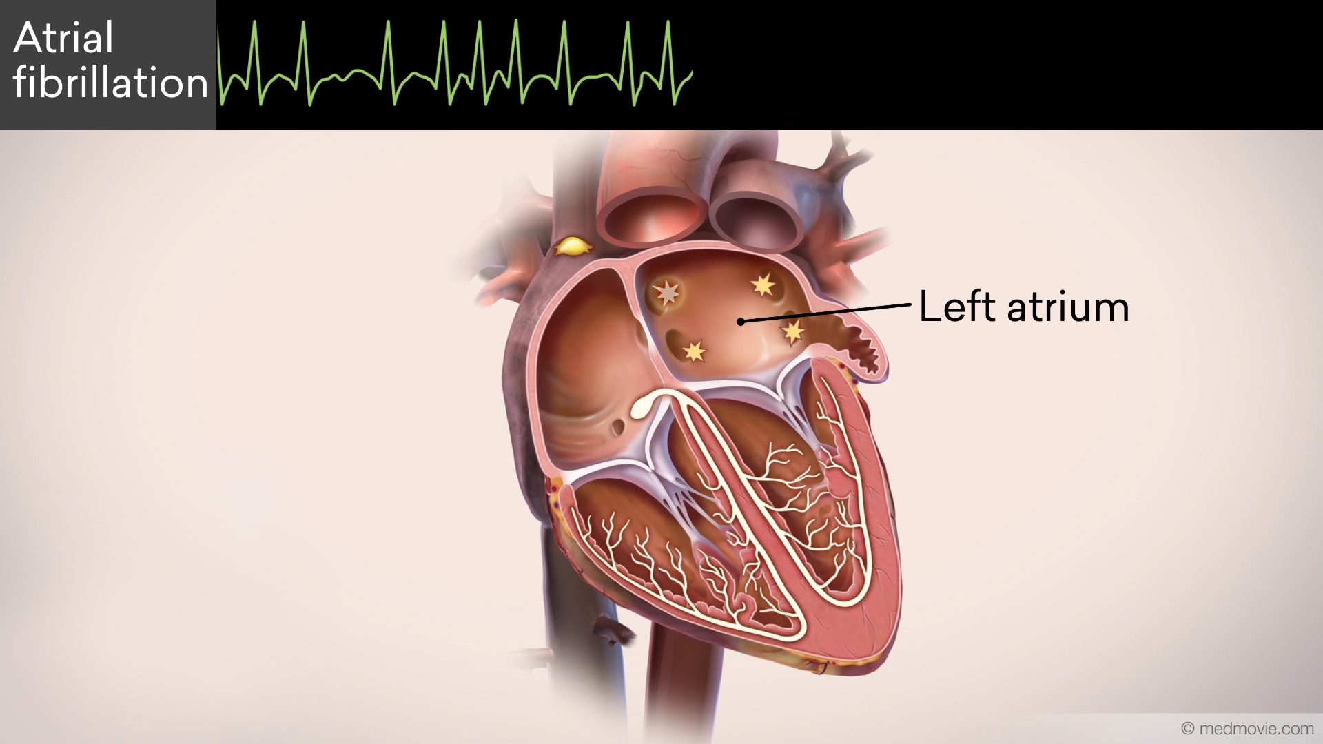 Atrial Fibrillation OverviewAtrial fibrillation is the most common heart rhythm disorder. In atrial fibrillation, abnormal signals from the left…
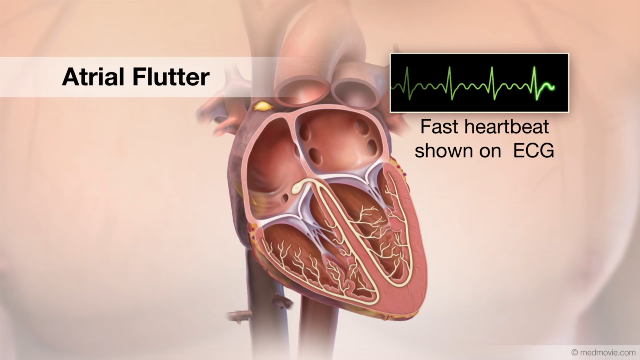 Atrial FlutterThe heartbeat is controlled by the electrical system of the heart. This system is made of several parts that tell the…
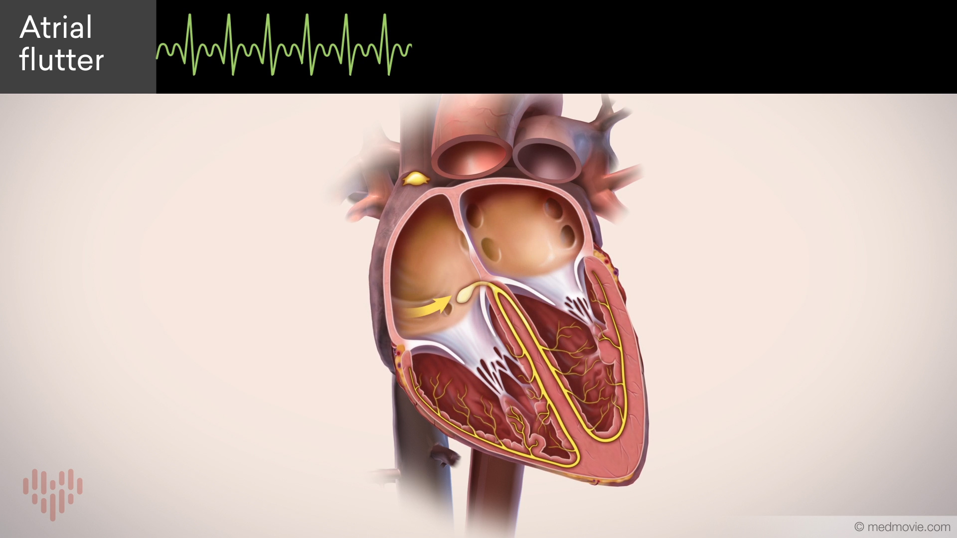 Atrial Flutter OverviewAtrial flutter is a common heart rhythm disorder. In atrial flutter, an abnormal electrical circuit spins in the right…Atrial Septal DefectAn atrial septal defect, or ASD, is a hole in the wall that separates the upper two chambers of the heart, known as the…
 Atrial TachycardiaThe heartbeat is controlled by the electrical system of the heart. This system is made up of several parts that tell…
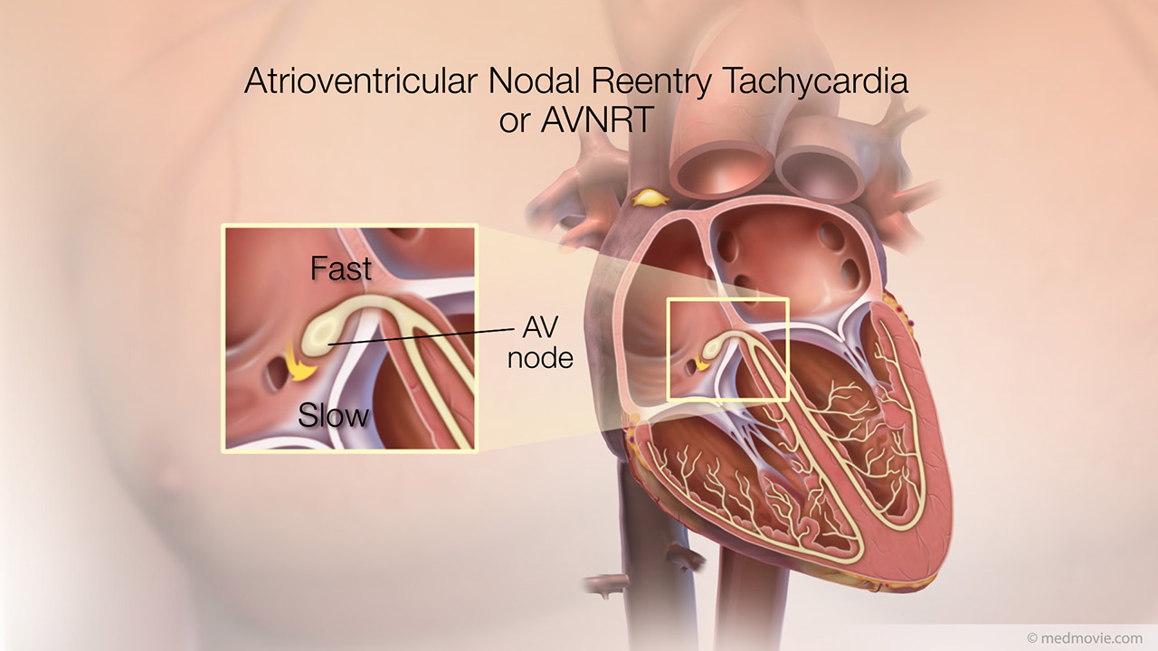 AVNRTThe heartbeat is controlled by the electrical system of the heart. This system is made up of several parts that tell…
 AVRTThe heartbeat is controlled by the electrical system of the heart. This system is made up of several parts that tell…AV CanalThe atrioventricular canal, or AV canal, is the structure that forms the center of the heart. During normal fetal…
 AV Node AblationAV node ablation is a procedure used to correct irregular heartbeats by destroying the tissue within the AV node. AV…
 BradycardiaThe heartbeat is controlled by the electrical system of the heart. This system is made of several parts that tell the…
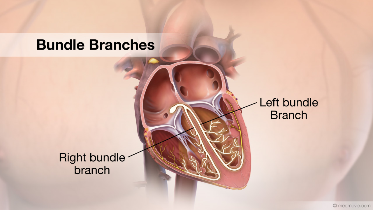 Bundle BranchesThe bundle branches are a part of the electrical system of the heart.
The electrical system controls the heartbeat…
Bundle BranchesThe bundle branches are a part of the electrical system of the heart.
The electrical system controls the heartbeat… Bundle of HisThe bundle of His is a part of the electrical system of the heart. It is a collection of cells that carry electrical…
 Cardiac CatheterizationCardiac catheterization refers to a procedure used to diagnose and treat certain heart conditions.
To initiate a…
Cardiac CatheterizationCardiac catheterization refers to a procedure used to diagnose and treat certain heart conditions.
To initiate a… CRT DeviceA cardiac resynchronization therapy (CRT) device is a battery-powered device that sends electrical signals to your…
 Catheter AblationThe electrical system of the heart controls each heartbeat. Electrical impulses generated by special tissue (nodes)…Coarctation of AortaCoarctation of the Aorta is a congenital defect in which the upper part of the descending aorta is severely narrowed or…
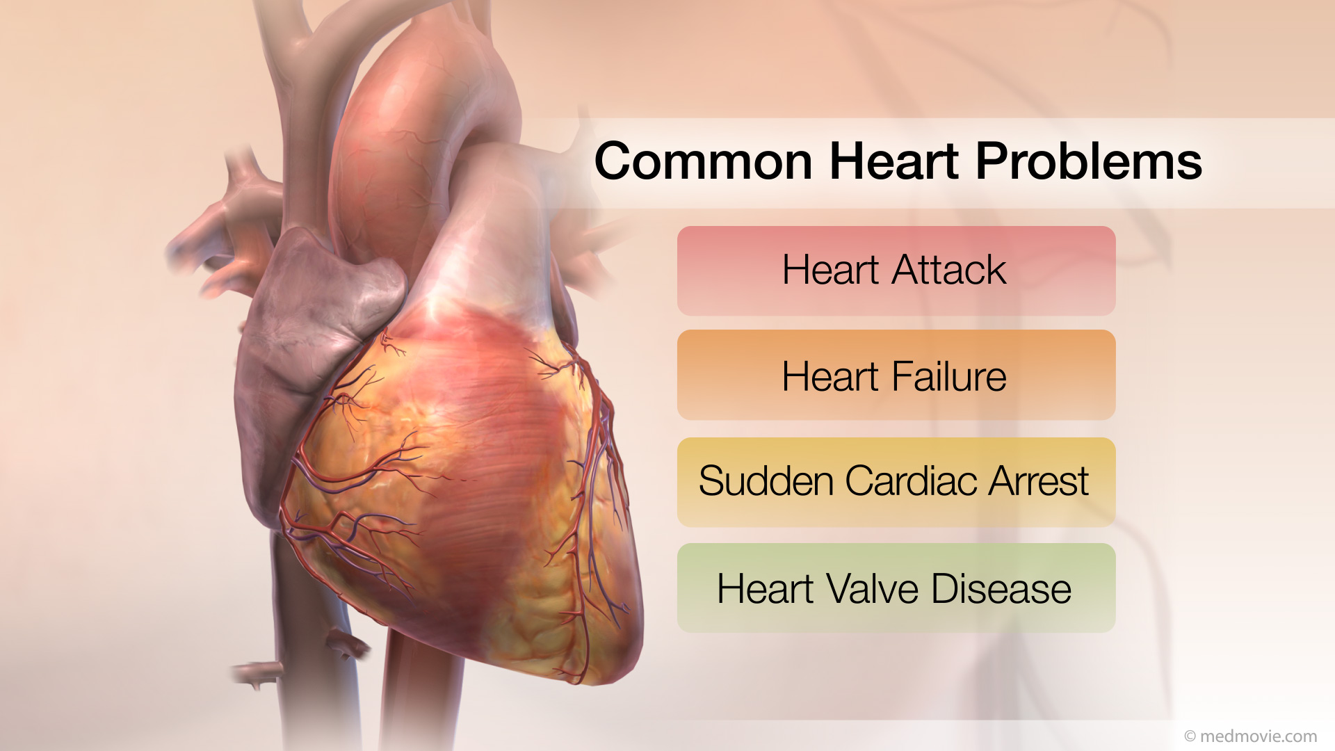 Common Heart ProblemsThe arteries, muscle, conductive tissue and valves of the heart work together to keep it beating normally. These four…
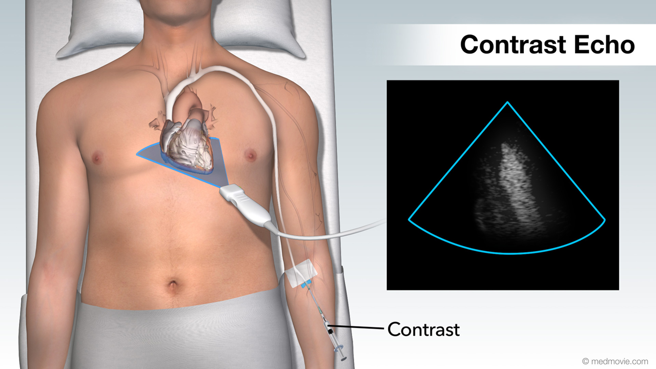 Contrast EchoHeart Ultrasound provides your doctor with moving images of your heart and takes excellent pictures that will help your…
 Coronary AngiogramA coronary angiogram is a test that uses x-ray imaging and contrast dye to view the coronary arteries, the blood…
 Coronary AngioplastyCoronary angioplasty (PTCA) is a procedure that opens up narrowed or blocked segments of the arteries that supply blood…
 Coronary Art. DiseaseCoronary artery disease is a condition in which the coronary arteries are narrowed by deposits called plaques.
The…
Coronary Art. DiseaseCoronary artery disease is a condition in which the coronary arteries are narrowed by deposits called plaques.
The… Coronary StentA Coronary Stent is a small expandable tube shaped device used to open narrowed coronary arteries that have interrupted…
 CRT OverviewA cardiac resynchronization therapy (CRT) device is a battery-powered device that sends electrical signals to your…
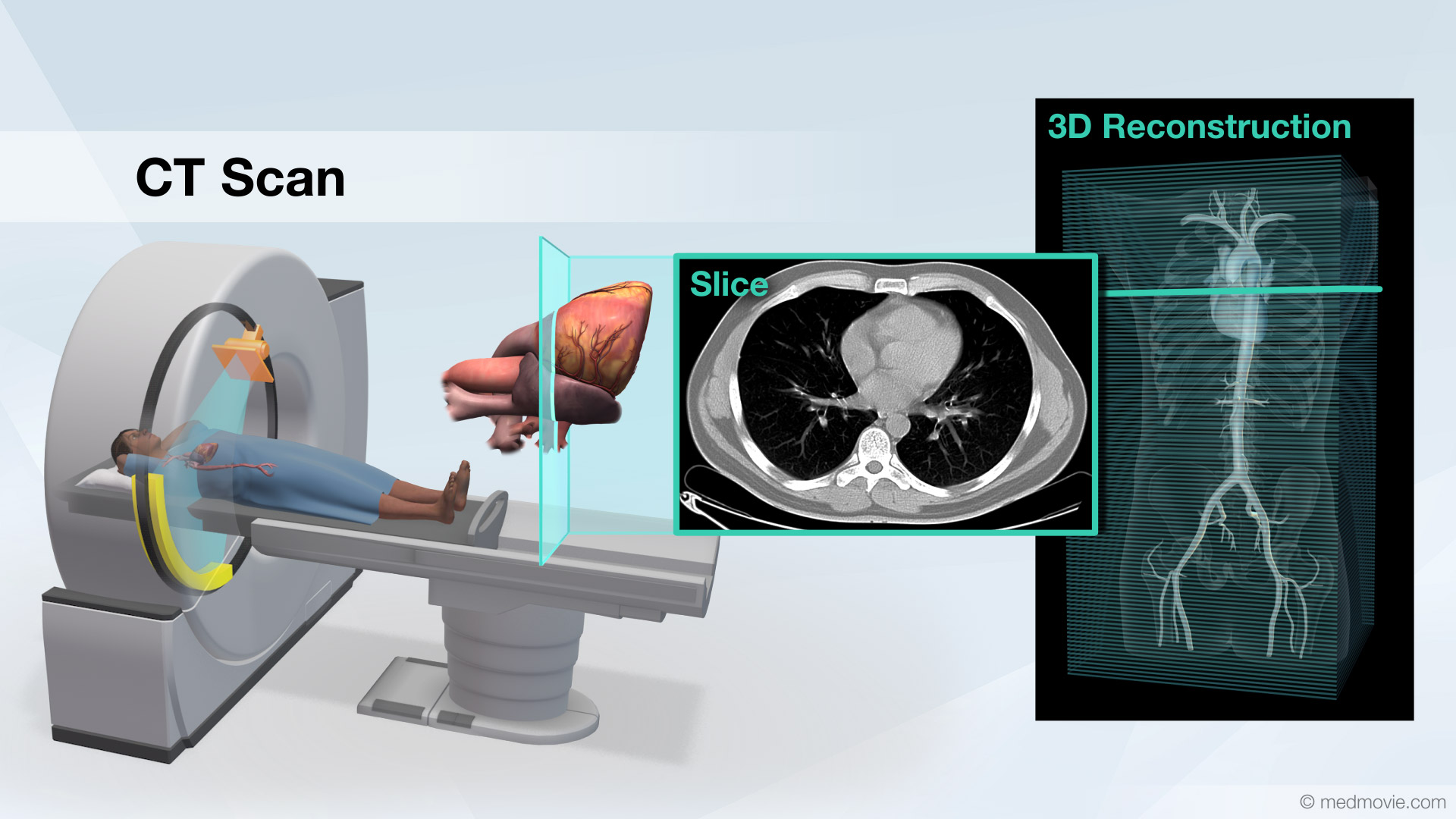 CT ScanA Computed Tomography scan, or CT scan, also known as a CAT scan, is a non-invasive diagnostic test that uses x-rays to…
 Dilated CardiomyopathyDilated cardiomyopathy is a term used to describe a condition of the heart in which one or more of the chambers of the…
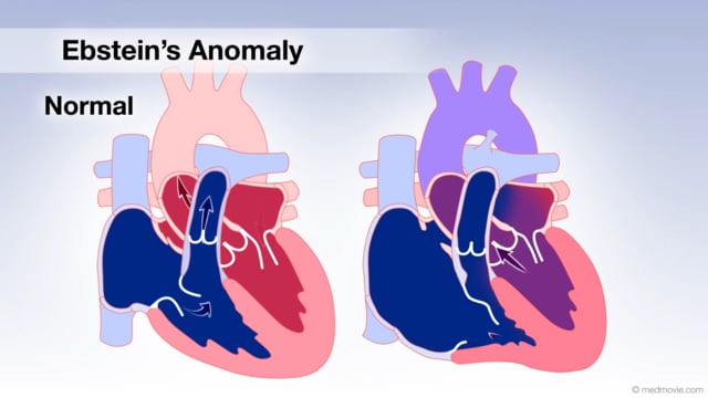 Ebstein's AnomalyThe tricuspid valve is the valve between the right atrium and the right ventricle, and in Ebstein’s anomaly it is…
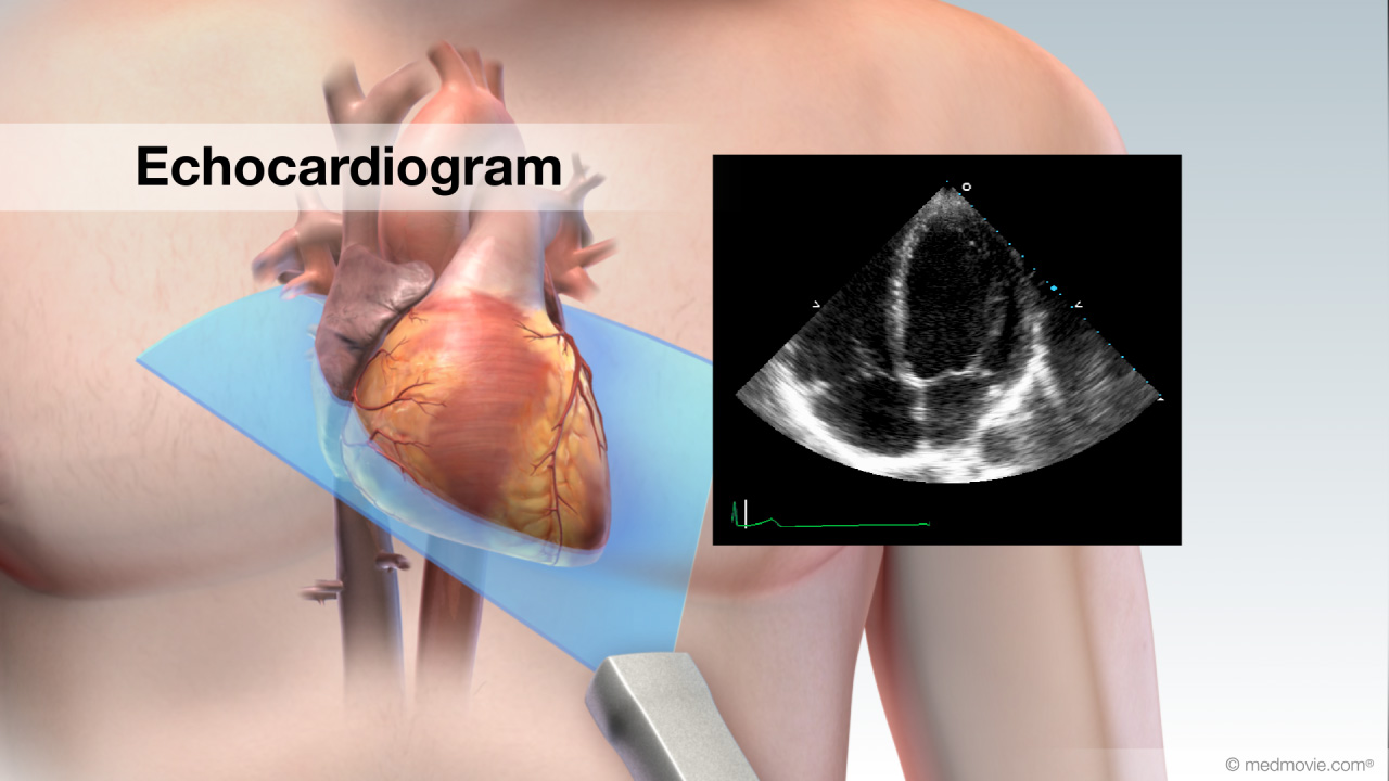 EchocardiogramAn echocardiogram, or cardiac ultrasound, is a diagnostic test that uses sound waves to view structures of the beating…
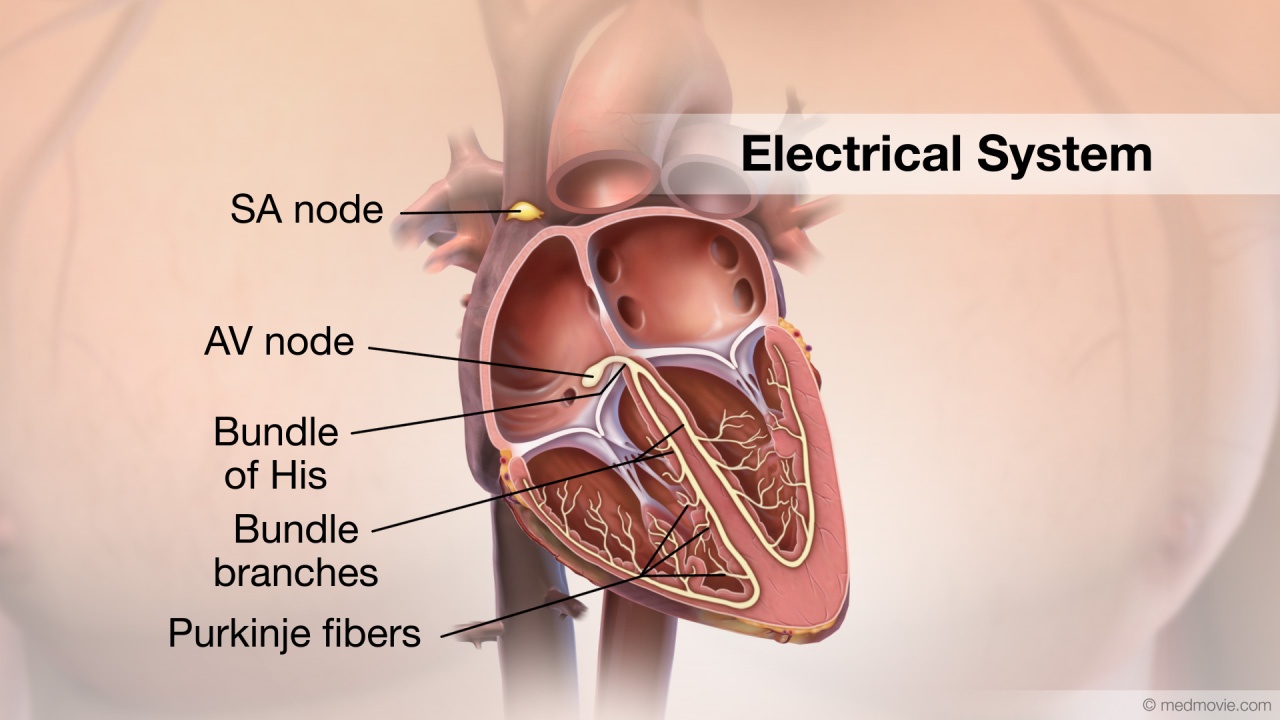 Electrical SystemThe purpose of the electrical system of the heart is to coordinate the pumping of the four chambers of the heart and to…
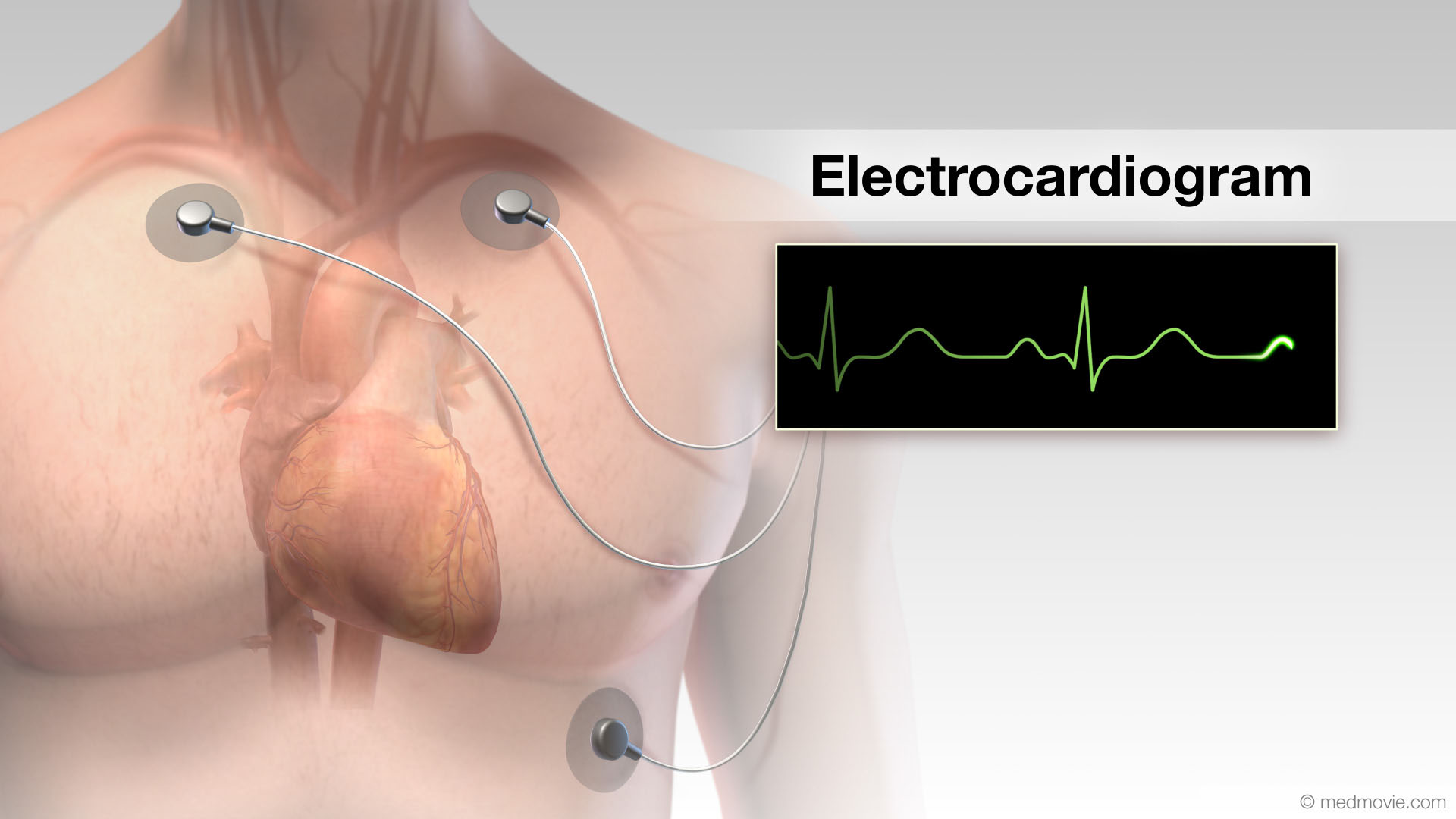 ElectrocardiogramThe electrocardiogram or ECG, records the electrical activity of the heart. It is used to determine if the heart has an…
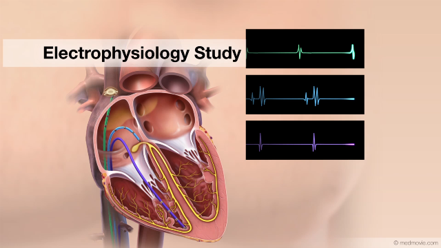 EP StudyAn electrophysiology study is performed to evaluate the electrical activity of the heart. The purpose of the electrical…
 Exercise Stress TestAn exercise stress test is a diagnostic test in which a person walks on a treadmill or pedals a stationary bicycle…
 Focal Atrial TachycardiaThe heartbeat is controlled by the electrical system of the heart. This system is made up of several parts that tell…
 Focal Ventricular TachycardiaThe heartbeat is controlled by the electrical system of the heart. This system is made up of several parts that tell…
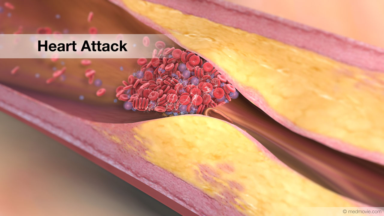 Heart AttackA heart attack happens when blood flow to an area of heart muscle suddenly becomes blocked and an area of heart muscle…
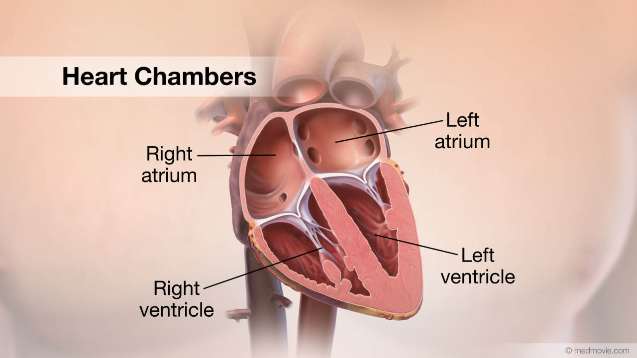 Heart ChambersThe heart is divided into four chambers. The two upper chambers are called the atria. The right atrium and left atrium…
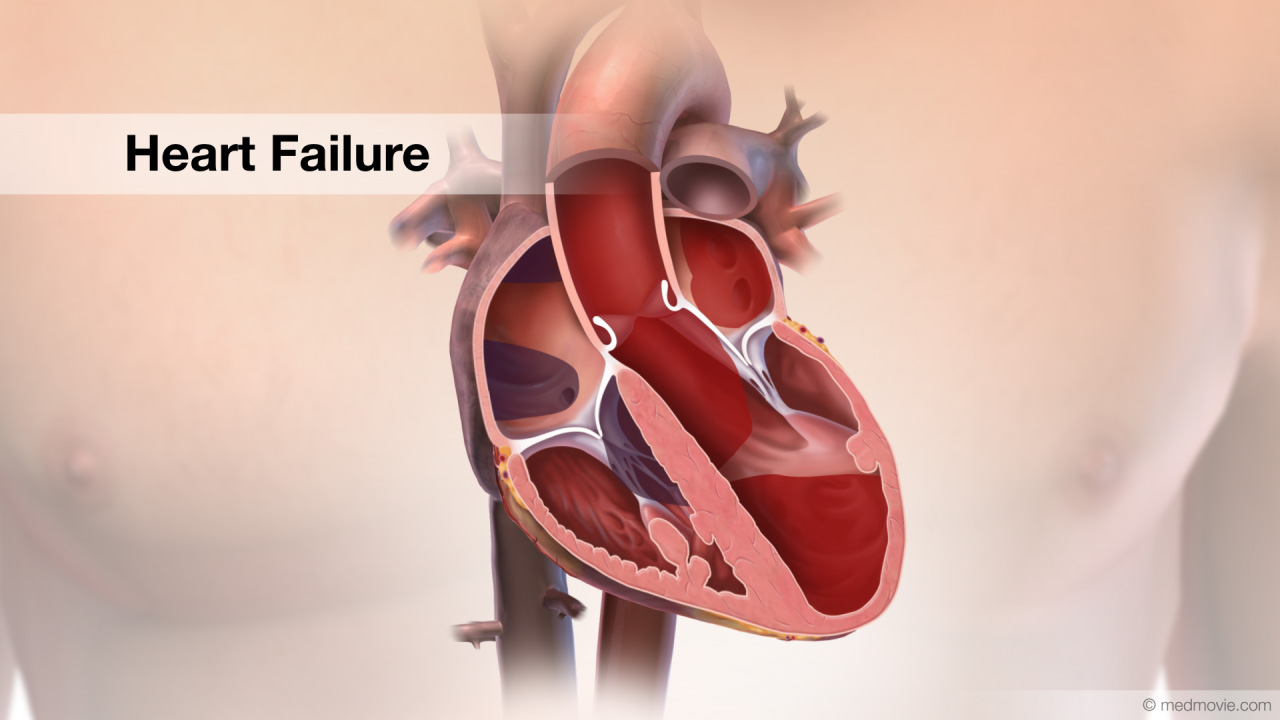 Heart FailureThe normal heart has strong muscular walls that pump blood out of the heart. Heart failure most often results from a…
 Heart Lung BypassA heart-lung -or cardiopulmonary bypass - machine is used during heart surgery to deliver oxygenated blood to the body…
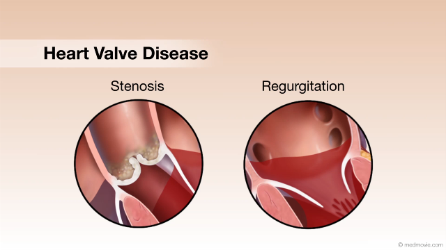 Heart Valve DiseaseValvular heart disease is any condition that disrupts the proper function of the valve. There are four valves in the…
 Heart Valve SurgeryHeart valve replacement surgery replaces an abnormal or diseased heart valve with a healthy one.
There are four…
Heart Valve SurgeryHeart valve replacement surgery replaces an abnormal or diseased heart valve with a healthy one.
There are four…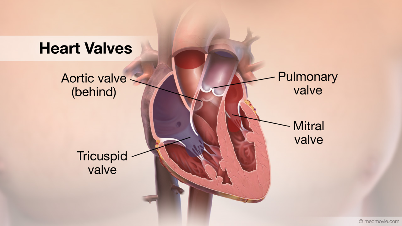 Heart ValvesThe heart has four valves. The mitral valve, the tricuspid valve, the pulmonary valve and the aortic valve. The mitral…
 Hidden RiskFeeling fine does not equal low risk for heart attacks. With coronary artery disease or “C”, “A”, “D” it’s easy to…
 High Blood PressureHigh blood pressure, or hypertension, is a condition in which blood pressure levels are measured above the normal…Hypertrophic CardiomyopathyThe normal heart has strong muscular walls that pump blood out of the heart.
Hypertrophic cardiomyopathy is a…
High Blood PressureHigh blood pressure, or hypertension, is a condition in which blood pressure levels are measured above the normal…Hypertrophic CardiomyopathyThe normal heart has strong muscular walls that pump blood out of the heart.
Hypertrophic cardiomyopathy is a… Hypoplastic Left Heart SyndromeHypoplastic left heart syndrome is the result of underdevelopment of the left heart structures and includes a tiny, or…
 ICD DeviceAn Implantable Cardioverter Defibrillator, or ICD, is a battery-powered device that keeps track of your heartbeat, and…
 ICD OverviewAn Implantable Cardioverter Defibrillator, or ICD, is a battery-powered device that keeps track of your heartbeat and…
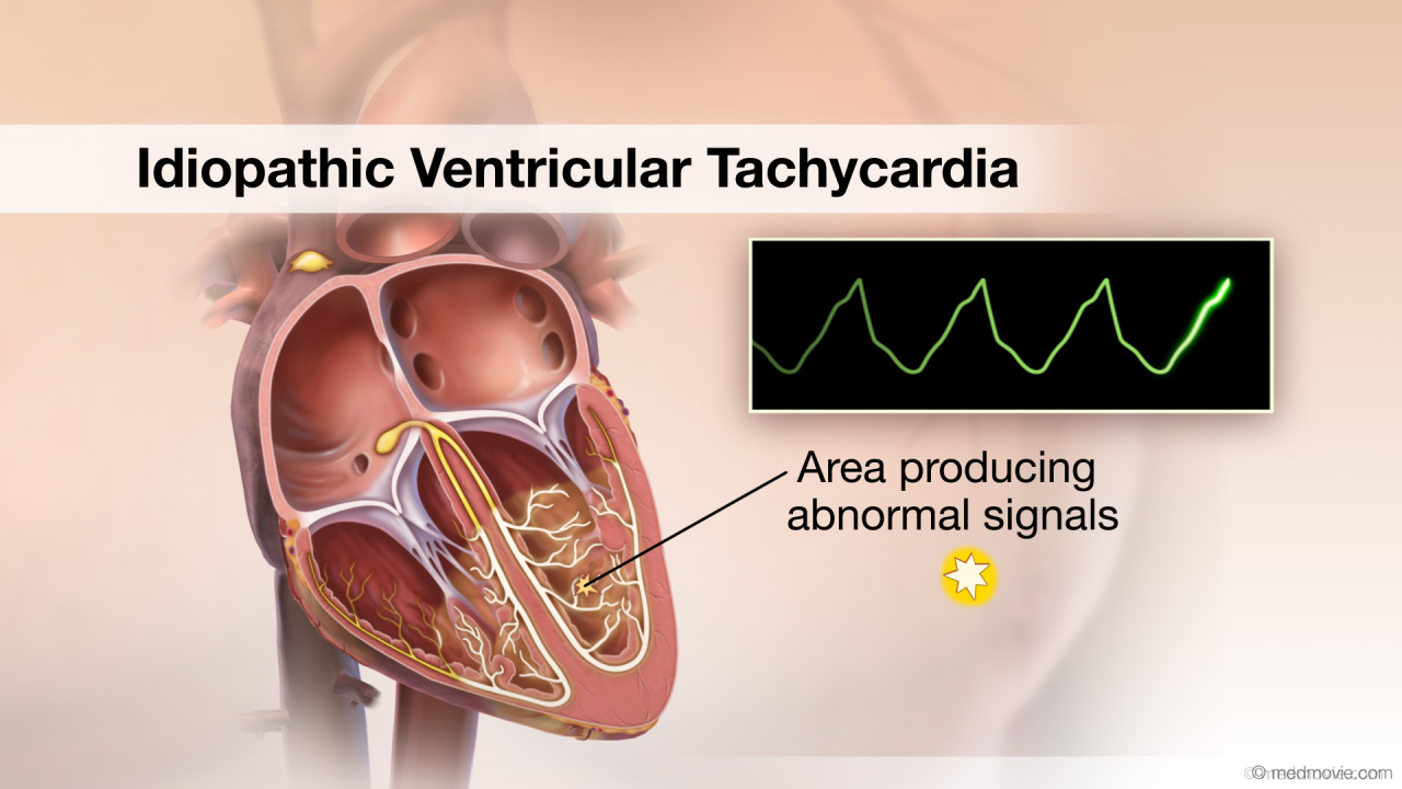 Idiopathic Ventricular TachycardiaThe heartbeat is controlled by the electrical system of the heart. This system is made up of several parts that tell…
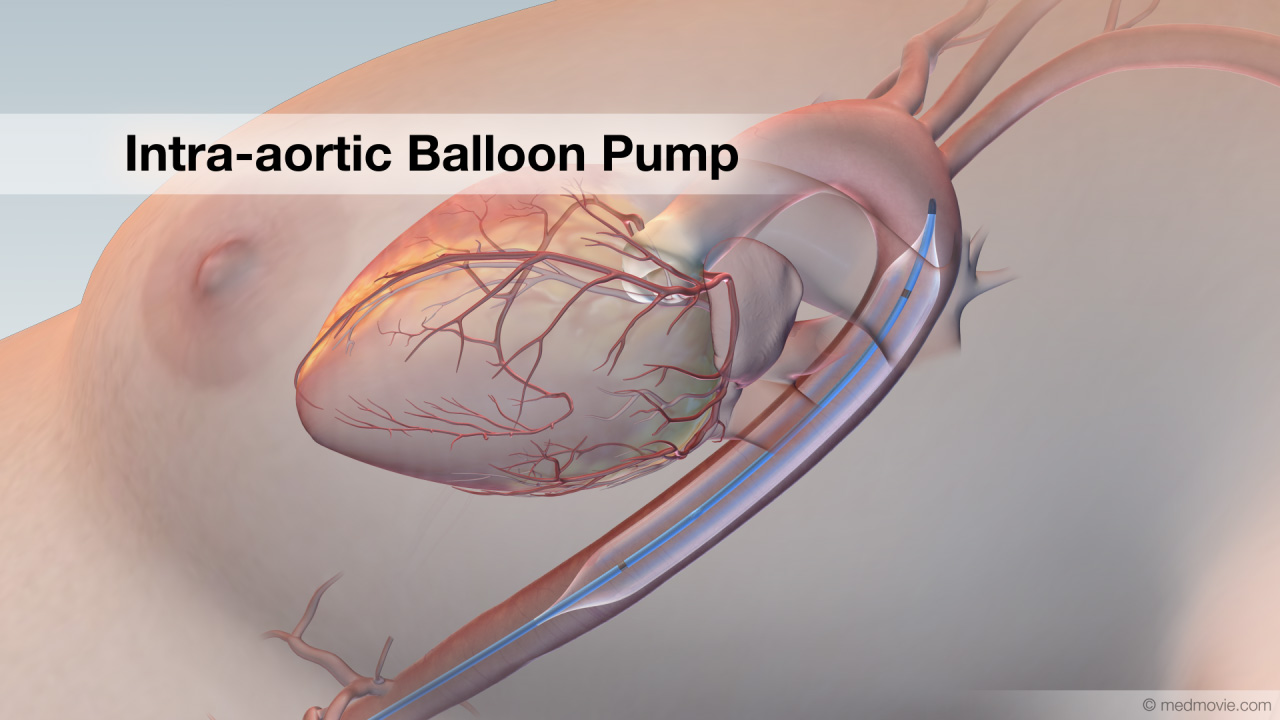 Intra-aortic BalloonAn intra-aortic balloon pump or IABP is an inflatable balloon that is surgically implanted into the aorta to help the…
 Ischemic Ventricular TachycardiaThe heartbeat is controlled by the electrical system of the heart. This system is made up of several parts that tell…
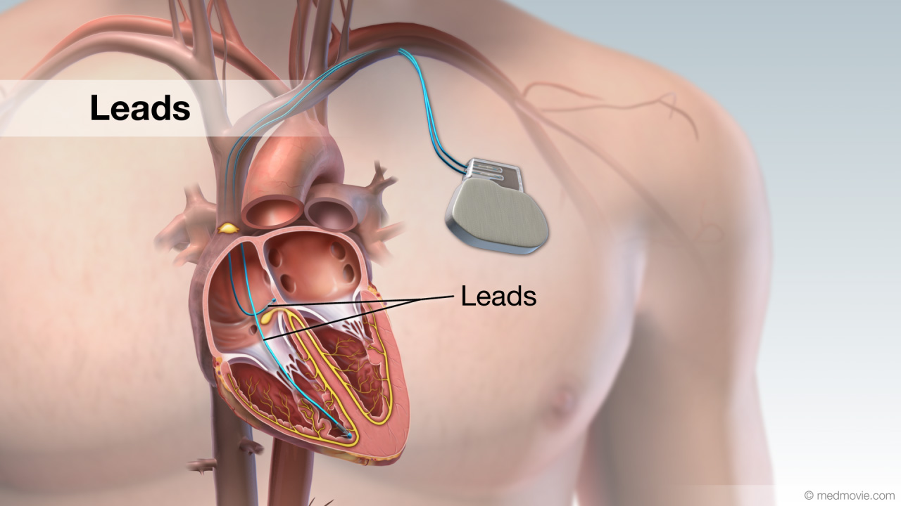 LeadA lead is a device that measures the passage of electrical signals. Leads are used in electrocardiograms to record the…
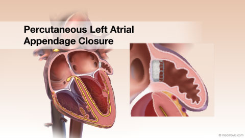 LAACA left atrial appendage closure device can be used for atrial fibrillation patients to help prevent strokes and as an…
 Long QT SyndromeLong QT Syndrome is a condition that is characterized by a delay in repolarization of the heart after the initial…
 Mitral Valve ClipA mitral valve clip is a device that supports an improperly functioning mitral valve.
Mitral valve leaflets that do…
Mitral Valve ClipA mitral valve clip is a device that supports an improperly functioning mitral valve.
Mitral valve leaflets that do…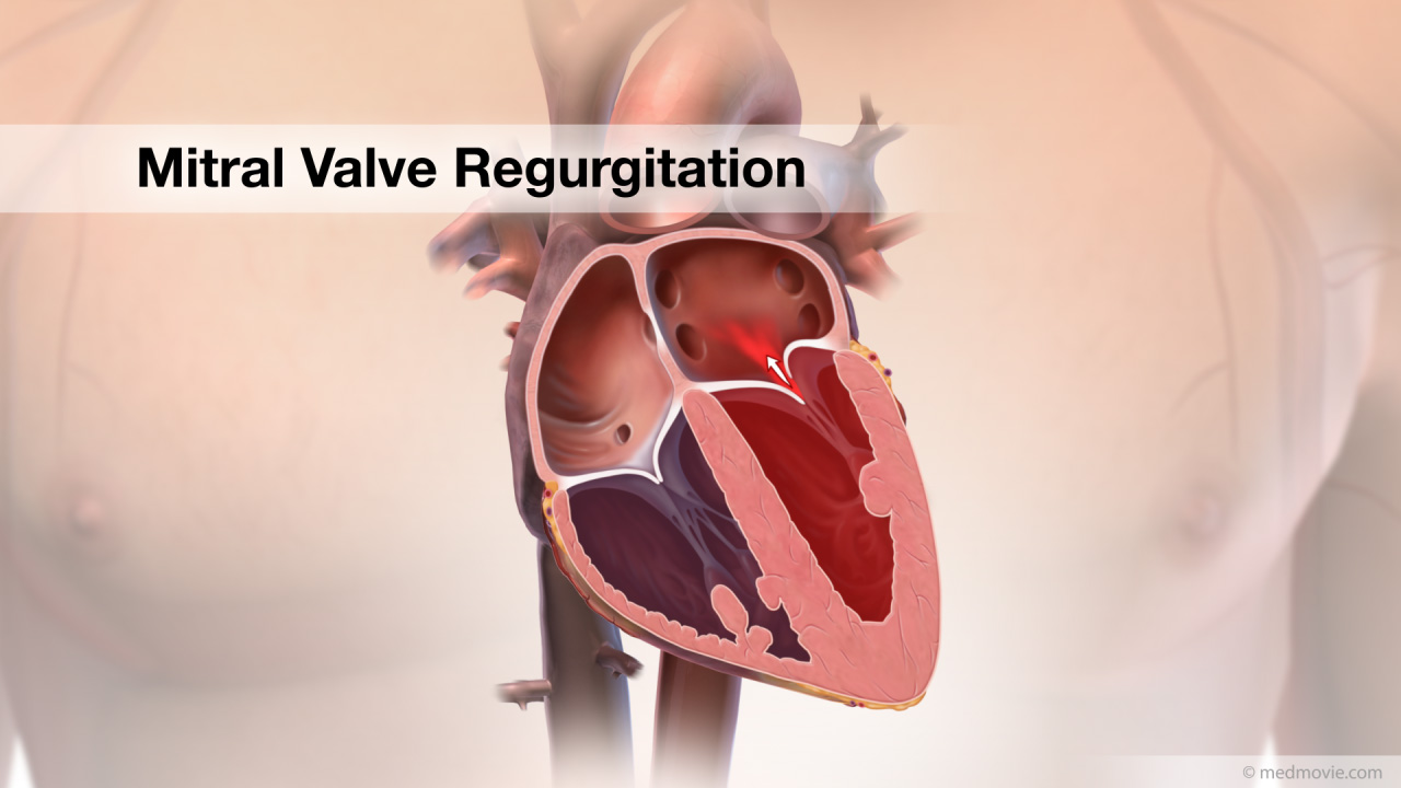 Mitral RegurgitationThe heart has four valves that regulate blood flow through the heart. The mitral valve is the heart valve located…
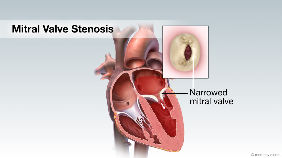 Mitral Valve StenosisMitral valve stenosis is a condition when the mitral valve does not open properly, narrowing the opening, reducing the…
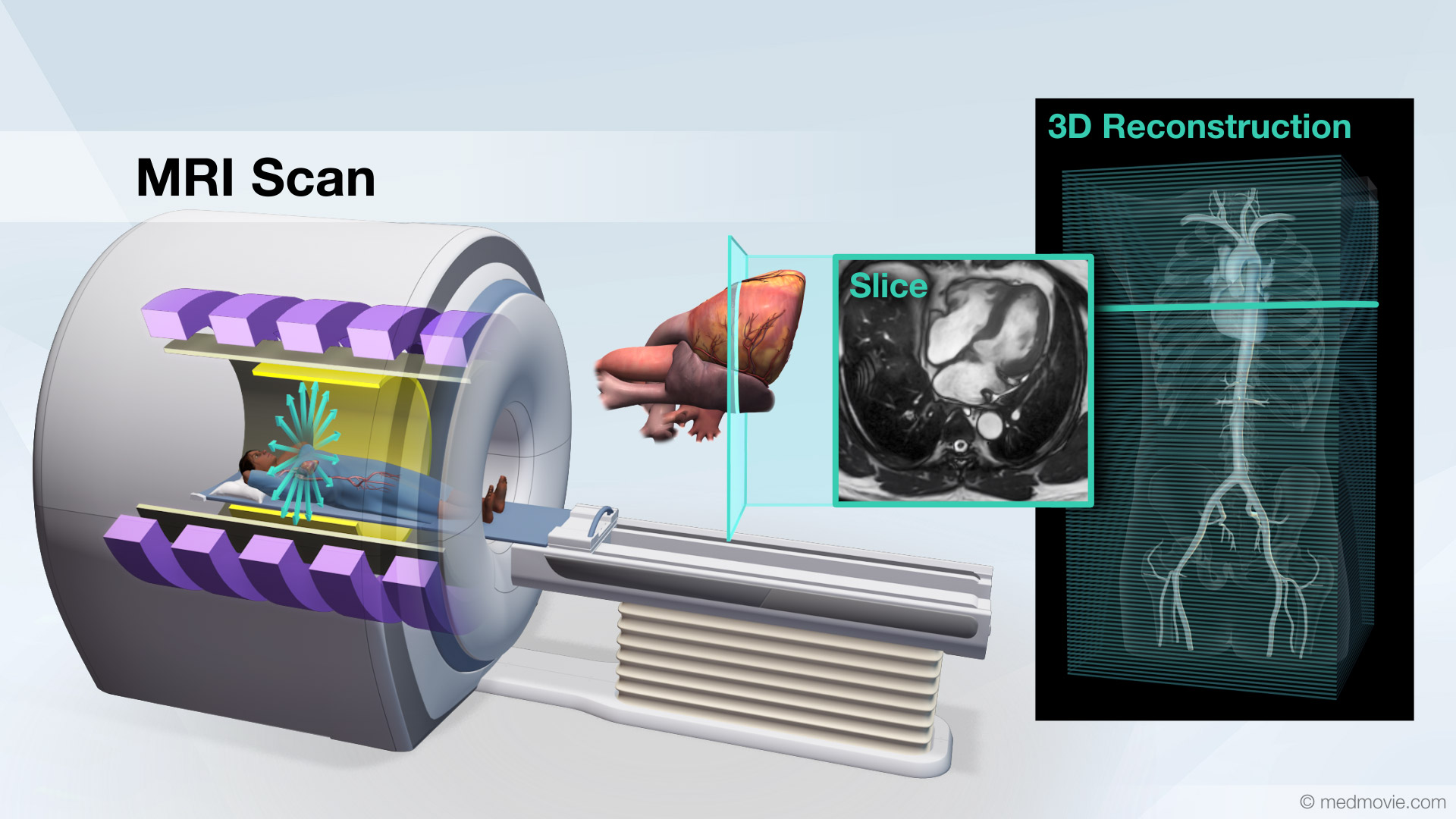 MRI ScanA Magnetic Resonance Imaging scan, or MRI scan, is a non-invasive diagnostic test that uses a magnetic field and radio…
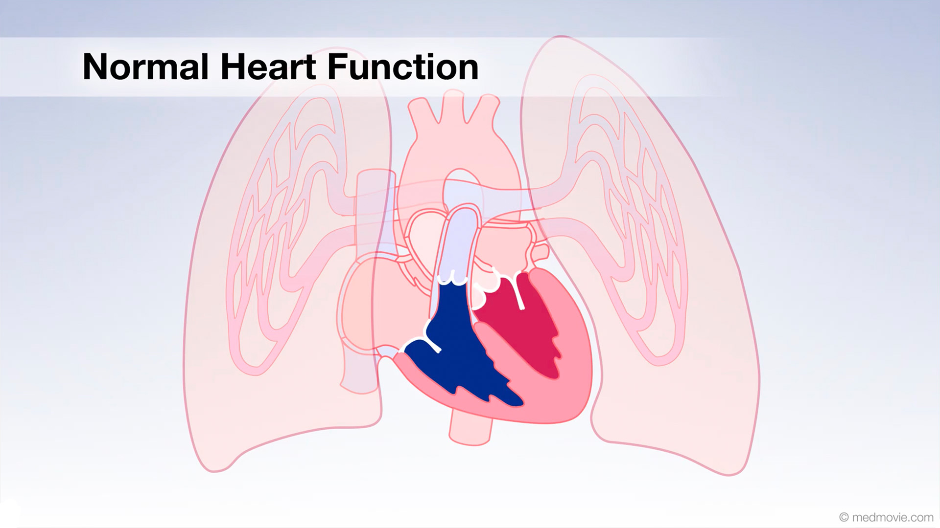 Normal Heart FunctionBlood flows through the heart in a specific pattern in order to pump deoxygenated, or blue blood to the lungs and…
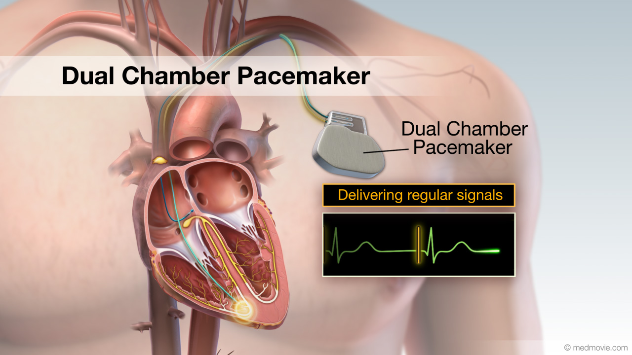 Pacemaker - Dual ChamberA pacemaker is a battery-powered device that sends electrical signals to your heart to help it beat at a proper rate or…
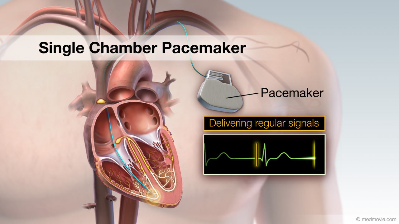 Pacemaker - Single ChamberA pacemaker is a battery-powered device that sends electrical signals to your heart to help it beat at a proper rate or…Patent Ductus Arter.The ductus arteriosus is a blood vessel that connects the blood vessel going to the lungs, known as the pulmonary…
 PET ScanA Positron Emission Tomography scan, or PET scan, is a nuclear medicine imaging technique that uses small amounts of a…
 Physical ActivityA sedentary lifestyle is one of the major risk factors for cardiovascular disease. A sedentary lifestyle is…
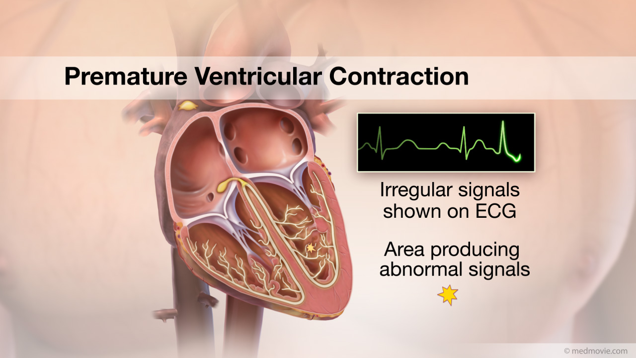 PVCThe heartbeat is controlled by the electrical system of the heart. This system is made up of several parts that tell…
 Pulm. Valve StenosisThe pulmonary valve is located between the right ventricle and the large blood vessel to the lungs which is called the…
 Quitting SmokingQuitting smoking is not easy, but there are things you can do to increase your chances of success. Planning strategies…
 RVOT Ventricular TachycardiaThe heartbeat is controlled by the electrical system of the heart. This system is made up of several parts that tell…
 Sinus RhythmA normal heartbeat produces a regular, identifiable pattern: the P wave, the QRS complex, and the T wave. These…
 Sinus TachycardiaThe heartbeat is controlled by the electrical system of the heart. This system is made of several parts that tell the…
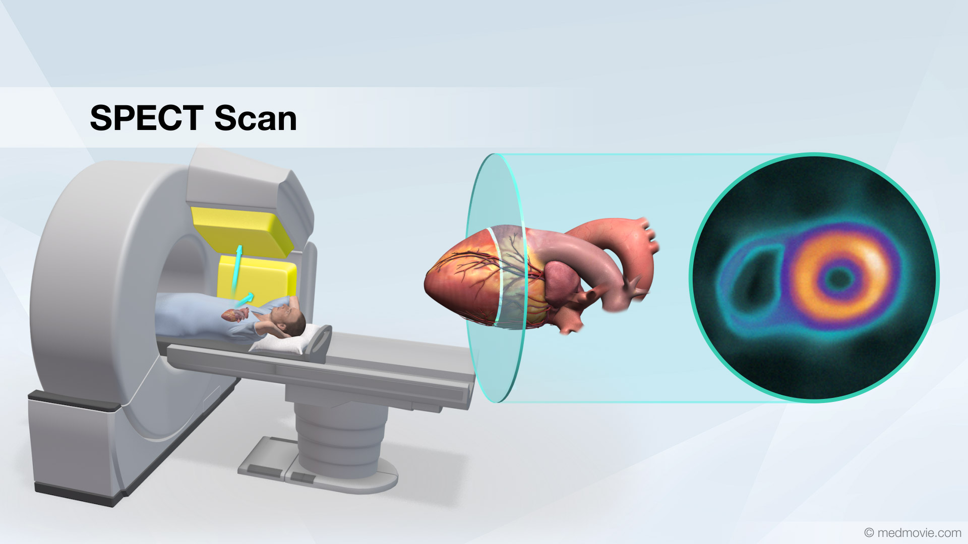 SPECT ScanA Single Photon Emission Computed Tomography scan, or SPECT scan, is a nuclear medicine imaging technique that uses…
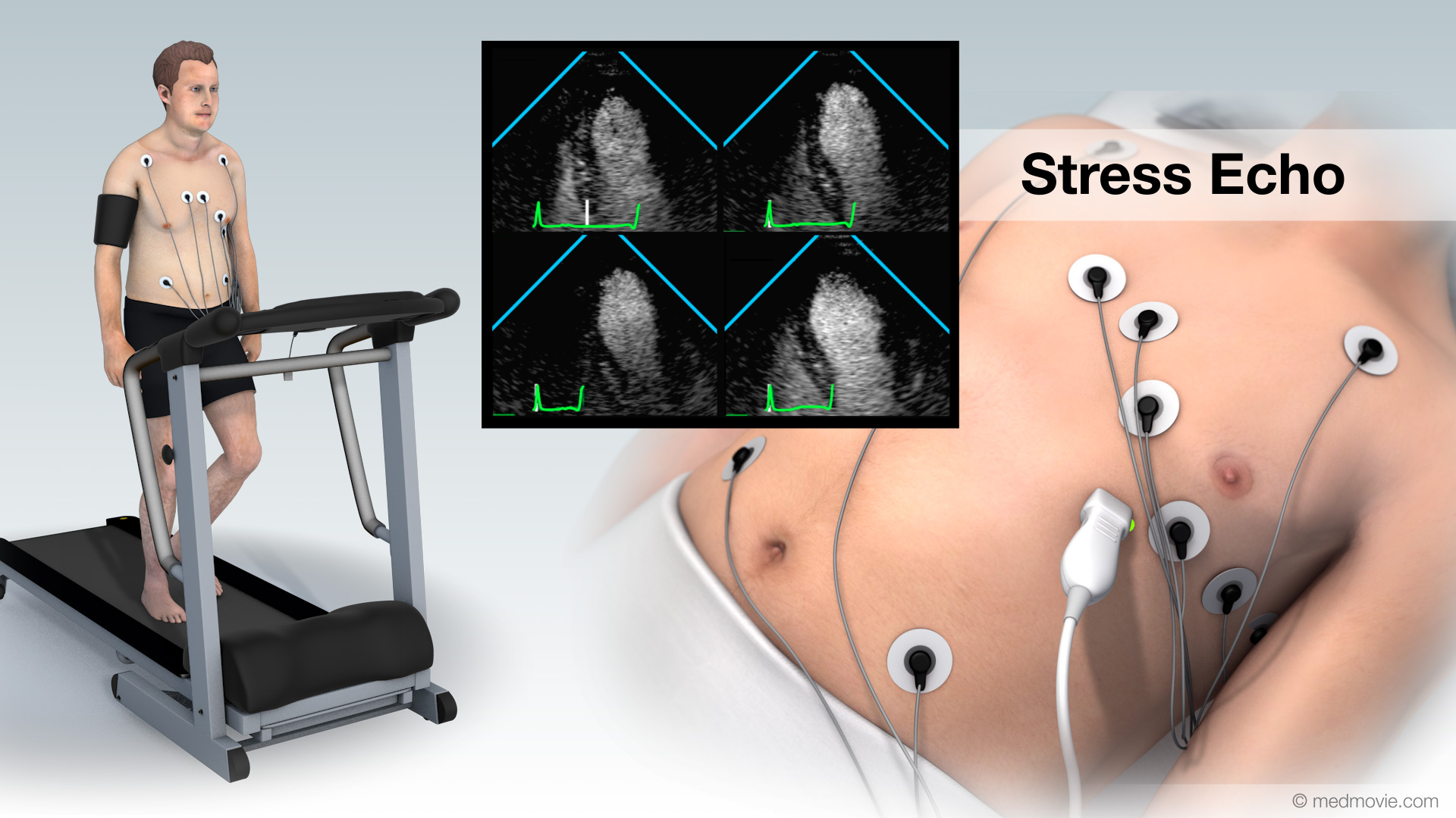 Stress EchoA stress echo, also called an exercise heart ultrasound is used to evaluate heart function by combining an exercise…
 Sudden Cardiac ArrestSudden cardiac arrest (SCA) is an event caused by a problem with the heart's "electrical" system. SCA occurs when the…
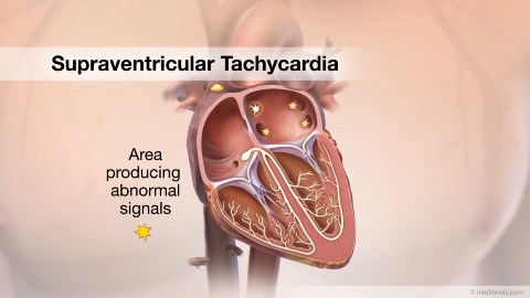 SVTThe heartbeat is controlled by the electrical system of the heart. This system is made up of several parts that tell…
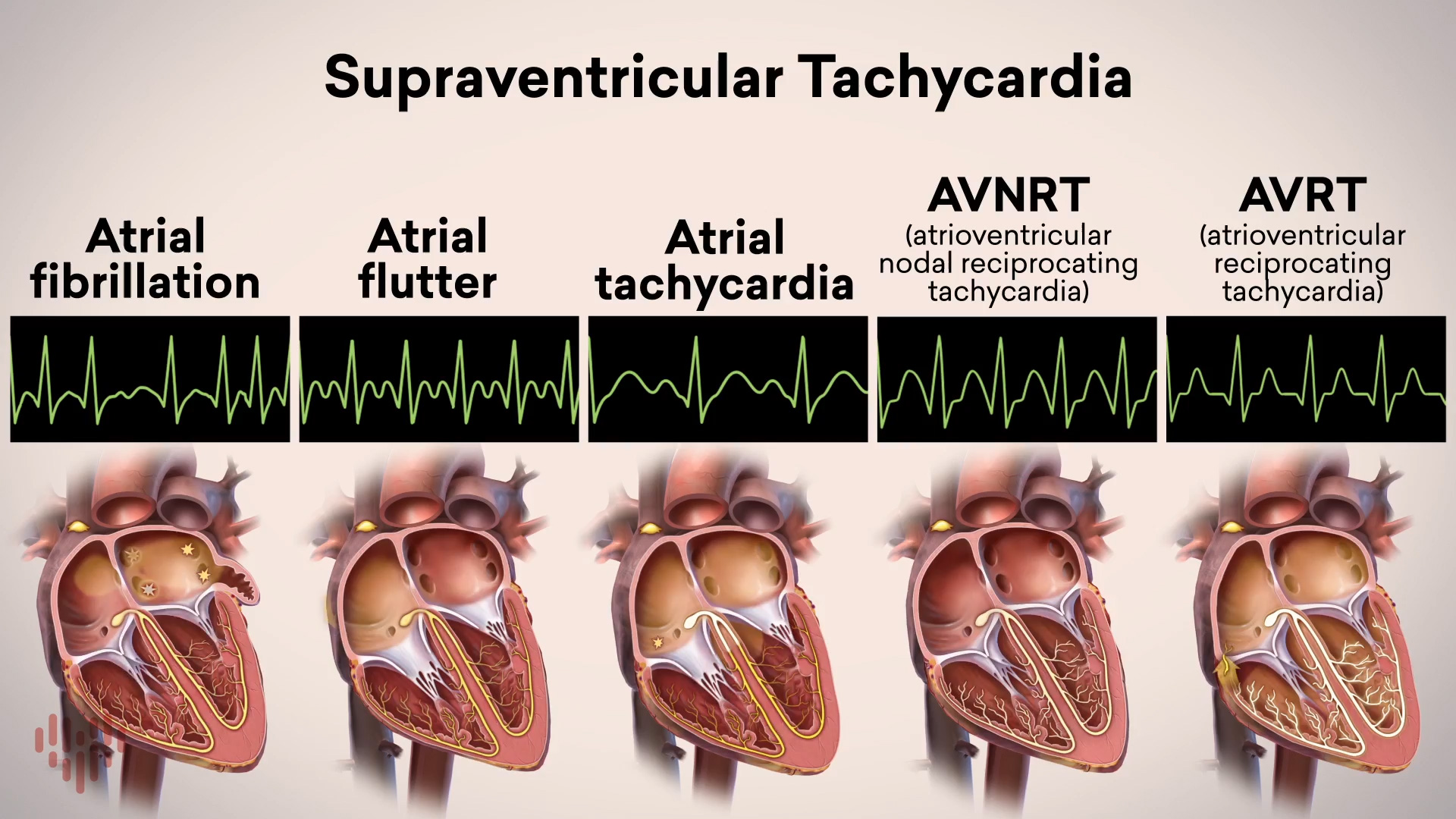 Supraventricular Tachycardia OverviewSupraventricular tachycardias are heart conditions with a fast heart rhythm and an electrical abnormality of the atria…
 Transcatheter Aortic Valve ReplacementTranscatheter aortic valve replacement, or TAVR, is a treatment for aortic valve stenosis, which is a narrowing of the…
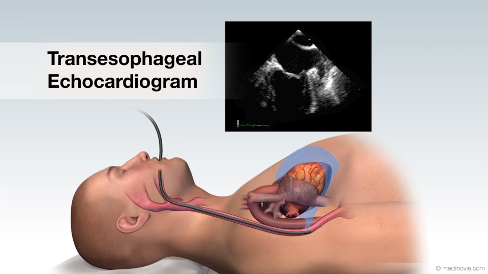 Transesophageal EchocardiogramA transesophageal echocardiogram is a diagnostic test used to view the structures of the beating heart. In this…
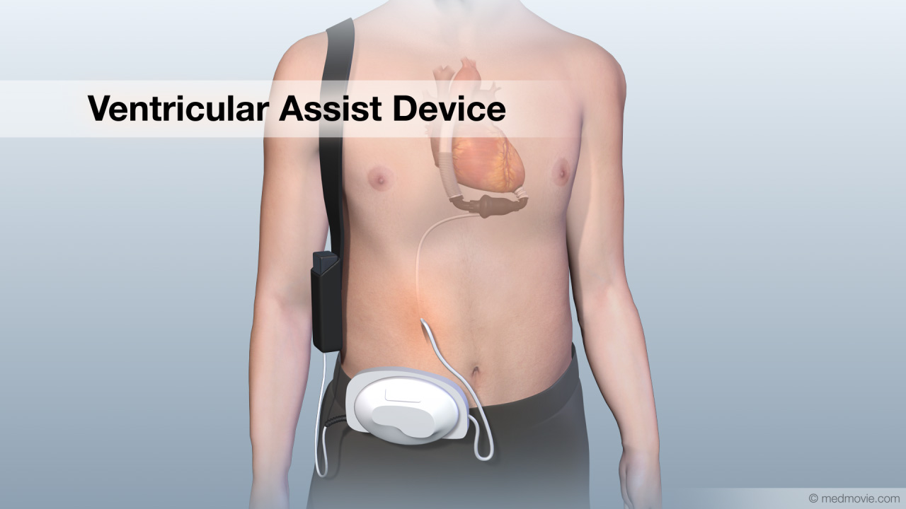 Ventricular Assist DeviceA ventricular assist device, VAD, is a pumping device that is used to help a heart that can no longer pump blood…
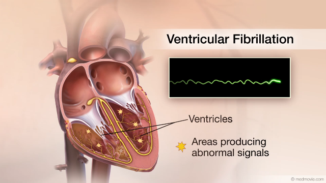 Ventricular FibrillationThe heartbeat is controlled by the electrical system of the heart. This system is made up of several parts that tell…
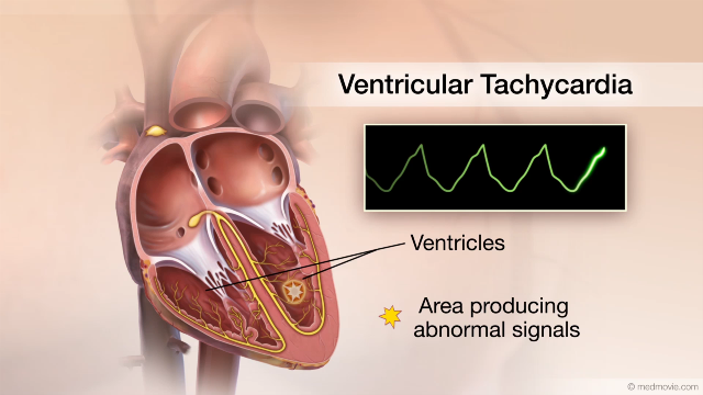 Ventric. TachycardiaThe heartbeat is controlled by the electrical system of the heart. This system is made of several parts that tell the…