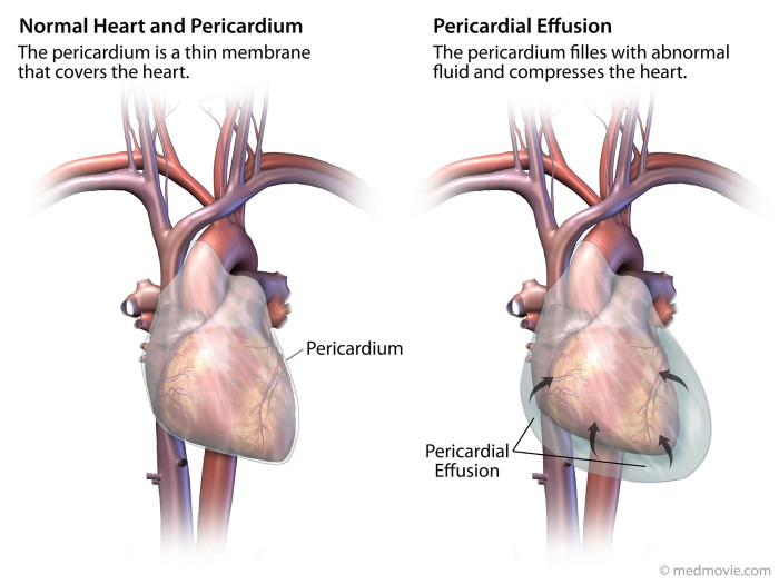
The first animation shows ultrasounds taken when the patient was initially admitted to the hospital. The ultrasounds show a large pericardial effusion and a right atrium that collapses with each beat. The pericardial effusion was later drained.
A printed panel was created to show the jury what pericardial effusion was.
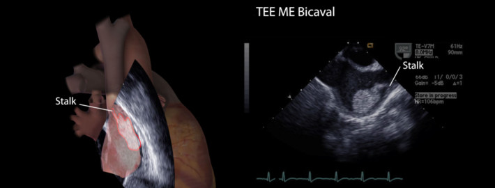
After four days, an additional ultrasound was performed. There was a mass in the superior right atrium. The physician presumed the mass was missed on the previous ultrasound, and thought that the mass was a pedunculated tumor. This animation shows that the previous exam views would have shown this mass if it existed 4 days prior. A tumor would have been excluded from possible diagnosis if the mass was determined to be absent in the earlier ultrasound.
This animation and printed panel show all the ultrasound views in which the mass could be clearly seen.
This video shows a reconstruction of the size and location of the mass, showing it was not insignificant enough to miss. The masses sudden appearance should have lead to the suspicion that the mass was a blood clot. Its location is also consistent with location of the atrial collapse which may have lead to myocardial damage or variations in blood flow.
The location of the atrial collapse may have lead to myocardial damage or abnormalities in blood flow which may have caused to the formation of a blood clot.
The mass’s location is consistent with location of the atrial collapse.
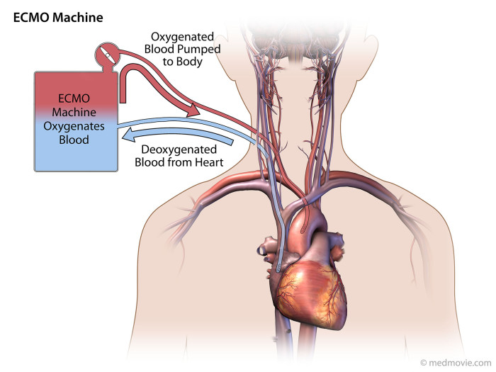
This misdiagnosis led to treating the mass as a tumor. The mass was biopsied which caused the clot to break loose and cause a pulmonary embolism. The facility did not have pediatric ECMO machine to maintain blood flow to the brain and body while surgery was performed to remove the clot. A printed panel was created to show the jury how a ECMO or heart lung bypass machine can provide oxygenated blood to the body and brain.